Hans Journal of Biomedicine
Vol.3 No.3(2013), Article ID:12054,5 pages DOI:10.12677/HJBM.2013.33004
Rapid Detection of Respiratory Bocavirus by Loop Mediated Isothermal Amplification
1College of Life Sciences, Hubei University, Wuhan
2Department of Laboratory Medicine, Wuhan Children’s Hospital, Wuhan
Email: *tangxingchun@hubu.edu.cn, *ahw066@126.com
Received: Apr. 16th, 2013; revised: Apr. 25th, 2013; accepted: May. 9th, 2013
Copyright © 2013 Zheming Yan et al. This is an open access article distributed under the Creative Commons Attribution License, which permits unrestricted use, distribution, and reproduction in any medium, provided the original work is properly cited.
ABSTRACT:
Objective: To Build a method (LAMP, Loop mediated isothermal amplification) for detecting Human Bocavirus. Methods: By designing 4 LAMP primers specified for 6 sites in NP1 gene sequence of Bocavirus, we built a system to detect Human Bocavirus. And we detected 60 oropharyngeal swab samples by using this system and realtime fluores-cence quantitative PCR to check whether this system works well at the same time. Results: We found that we can get the production through the LAMP amplification and the gel electrophoresis results of the 60 samples, in which we found 8 positive samples, are consensus between the realtime fluores-cence quantitative PCR and the LAMP system. Conclusion: LAMP, a kind of quick, simple, specific, sensitive and low cost method in detecting the nucleic acids of virus, is an ideal alternative for quick screening assay and clinical detection in medical institutions because of the experimental instruments and requirements are not high.
Keywords: LAMP—Loop Mediated Isothermal Amplification; Human Bocavirus; Realtime Fluores-Cence Quantitative PCR
环介导等温扩增技术快速检测呼吸道博卡病毒
闫明哲1,向 贇2,胡适可1,张家云2,汤行春1*,艾洪武2*
1湖北大学,生命科学学院,武汉
2武汉市儿童医院,检验科,武汉
Email: *tangxingchun@hubu.edu.cn, *ahw066@126.com
摘 要:
目的:建立一种检测呼吸道博卡病毒的环介导等温扩增方法(LAMP)。方法:通过博卡病毒NP1基因特异性序列的6个位点设计4条LAMP引物,建立LAMP检测体系。并对60例疑感染者的咽试子,同时采用实时荧光定量PCR和LAMP方法进行博卡病毒核酸的检测。结果:通过凝胶电泳能观察到LAMP方法扩增引物的存在,且采用此体系的检测60份咽试子中8份阳性反应和实时荧光定量PCR检测结果一致。结论:博卡病毒核酸的LAMP检测方法快速、简单、特异、灵敏、成本低,对实验仪器设备要求不高,适用于基层医疗机构临床检测和现场快速诊断。
收稿日期:2013年4月16日;修回日期:2013年4月25日;录用日期:2013年5月9日
关键词:环介导等温扩增技术;博卡病毒;实时荧光定量PCR
1. 引言
人博卡病毒(human bocavirus, HBoV)是2005年瑞典科学家Allander等[1]对呼吸道感染患儿鼻咽分泌物进行检测时,在鼻咽分泌物中发现的一种新病毒。HBoV属于细小病毒科(parvoviridae),细小病毒亚科,博卡病毒属,是单链DNA病毒,会引起急性呼吸道感染。HBoV在儿童中,特别是<3岁的婴幼儿有很高的感染率以及共感染,但HBoV在成人中很少检测到[2,3]。环介导等温扩增技术(Loop Mediated Isothermal Amplification,LAMP),是指在一种具有链置换活性的DNA聚合酶的协助下使用2对引物对模板的6个位点在恒温(60℃~65℃之间)条件下,1 h内即可完成反应[4]。目前LAMP已经被广泛的应用于各种样品的DNA和RNA检测中。Poon等利用LAMP技术建立了严重急性呼吸综合症冠状病毒(SARS-CoV)的检测方法[5]。也有人将此方法用于胚胎性别鉴定和不同病原体的检测[6]。
因此,本文建立了一种操作简便、灵敏度较高以及特异性较强的检测HBoV的LAMP方法。
2. 材料与方法
2.1. 标本的采集
根据“艾滋病和病毒性肝炎等重大传染病防治”科技重大专项传染病监测技术平台项目中的“发热呼吸道症候群监测方案”进行操作。收集2012年5~7月武汉市儿童医院检测儿童的咽试子。将咽试子样本加入一定量缓冲液PBS充分振荡,然后8000 rpm离心5 min,吸取上清液加入到冻存管中保存到−80℃备用。
2.2. 仪器与试剂
仪器:PCR仪器(applied biosystems),核酸提取试剂盒(Roche),LighCycler 实时荧光定量RT-PCR仪器(Roche),电泳仪(Biometra)等。
试剂:BstDNA聚合酶、MgSO4、10×Buffer、MgCl2、dNTP、Taq酶、SYBER Green I等。
2.3. HBoV病毒DNA提取和HBoV病毒 实时荧光定量PCR检测
用核酸提取试剂盒提取DNA[7]。再通过以下程序进行实时荧光定量RT-PCR扩增,Pre-Incubation:95℃(5 min),1个循环;Amplification:95℃(10 s),55℃(15 s),72℃(10 s),40个循环;Melting Curve:95℃(1 min),65℃(10 s),1个循环;Cooling:40℃(10 s), 1个循环。
正向引物:5’-GGCTCCTGCTCTAGGAAATAAA GAG-3’
反向引物:5’-CCTGCTGTTAGGTCGTTGTTGTA T GT-3’
2.4. LAMP引物的设计
通过在线的LAMP引物设计软件Primer Explorer V4设计2对引物,包括1对外引物(F3和B3)、1对内引物(FIP和BIP),其中要注意FIC和BIC的5’端ΔG的绝对值要>4,其它的3’端ΔG值 > 4;引物在设计时使其GC含量介于40%~65%之间,但是当引物的GC含量介于50%~60%时,引物的质量相对较好;F2区段的5’端到B2区段的5’端(LAMP反应扩增的区域)之间的距离建议是120~180 bp;F3区段的3’端到F2区段的5’端之间的距离是0~20 bp。同理B2和B3之间的距离是0~20 bp。F2区段的5’端到F1区段的5’端之间的距离是40~60 bp[8]。
外引物:
F3 5’-ATCAGCCACCTATCGTCT-3’
B3 5’-CAATGCGAGTAGAGTGCC-3’
内引物:
FIP 5’-ATTGTCTTTTTTCCCCGATGTACTCCAC TGCTTCGAAGACCTC-3’
BIP 5’-TCCATACACTGTATTCAGTCAACACAG TAGAACCCACACCACC-3’
2.5. LAMP检测方法的建立
LAMP的反应体系中包括:内引物、外引物、Bst聚合酶、Thermopol Buffer、MgSO4、dNTPs、DEPC水、模板、MLV酶。其中要对内外引物浓度的比值、MgSO4浓度、dNTPs浓度、Thermopol Buffer浓度和反应时间以及反应温度对LAMP反应的影响进行优化试验,获得最好的反应条件,从而建立HBoV的LAMP检测体系。
2.6. LAMP扩增产物的电泳检测和染料检测
取6 μL LAMP扩增产物,于2%琼脂糖凝胶上进行电泳检测。扩增反应结束后,加入0.1 μL的10,000 × SYBR Green I染料,观察LAMP反应是否发生颜色变化。
2.7. LAMP检测HBoV的灵敏度
选取一份通过实时荧光定量PCR检测出来的阳性样本,提取核酸后进行常规PCR,电泳检测后回收目的条带。经浓度检测后进行10倍递进稀释,得到1.0 × 10⁹~1.0 × 10⁻¹ copies/μL的共11个稀释度,并用SYBR Green I染料进行检测LAMP扩增结果。
2.8. LAMP的特异性检测
提取甲型流感病毒、副流感病毒、呼吸道合胞病毒以及腺病毒的病毒核酸,按照所建立的HBoV检测方法进行特异性试验,以ddH2O作为阴性对照,待反应结束后于各反应试管中加入0.1 μL SYBR Green I观察颜色变化。
2.9. 博卡病毒临床样本的LAMP检测
从武汉市儿童医院选取60份儿童咽试子,分批次进行核酸提取,然后分别进行实时荧光定量PCR和LAMP检测。
3. 实验结果
3.1. LAMP检测方法的建立
通过对内外引物浓度的比值、MgSO4浓度、dNTPs浓度、Thermopol Buffer浓度和反应时间以及反应温度对LAMP的影响,确定最佳反应体系为 25 μL:Bst链置换聚合酶8 U(1 μL)、模板(2 μL)、DEPC水(9.5 μL)、10 × ThermopoI(2.5 μL)、MgSO4 10 mM(1.5 μL)、dNTPs 1.5 mM(3.5 μL)、F3/B3(1 μL/1 μL)、FIP/BIP(1 μL/1 μL)、MLV酶 10 U(1 μL)。反应条件为:65℃ 1 h;80℃ 3 min[8-10]。
3.1.1. MgSO4浓度优化
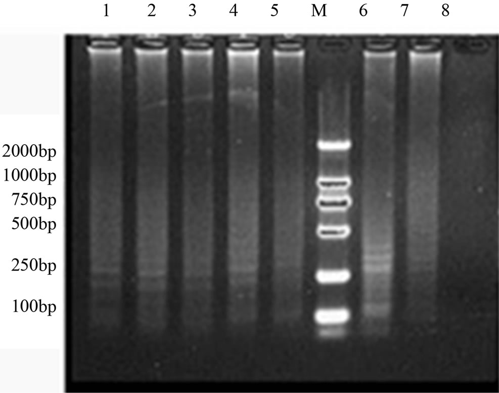
图1表明,MgSO4浓度在8~12 mM范围时,LAMP扩增效率无明显差异;4 mM浓度无LAMP反应发生,6 mM浓度时扩增效率较低。因此,最终确定MgSO4的最佳浓度为10 mM。
3.1.2. dNTPs浓度优化
图2表明,dNTPs浓度在1.5~2.0 mM时,LAMP扩增效率无明显差异。当dNTPs浓度为0.5 mM时,无LAMP扩增反应;dNTPs浓度为1.0 mM时,浓度过低,LAMP反应微弱,而dNTPs浓度为2.5 mM时,浓度过高会阻碍LAMP扩增效率,最终选择1.5 mM作为本试验的最佳dNTP反应浓度。
3.1.3. Bst DNA链置换聚合酶浓度优化
图3表明,Bst DNA链置换聚合酶浓度在4~10 U范围时,均有LAMP反应发生。而4 U的酶浓度扩增效率低,最终选择的最佳酶浓度为8 U。
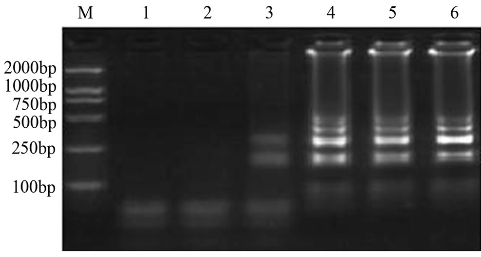
M: DL2000 Marker;1. ddH2O; 2. 4 mM; 3. 6 mM; 4. 8 mM; 5. 10 mM; 6. 12 mM.
Figure 1. The optimized results of MgSO4 concentration in LAMP reaction
图1. LAMP反应MgSO4浓度优化结果
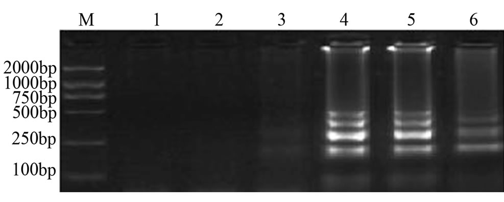
M: DL2000 Marker; 1. ddH2O; 2. 0.5 mM; 3. 1 mM; 4. 1.5 mM; 5. 2 mM; 6. 2.5 mM
Figure 2. The optimized results of dNTP concentration in LAMP reaction
图2. LAMP反应dNTPs浓度优化结果
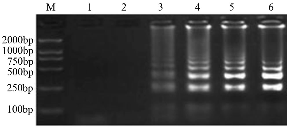
M: DL2000 Marker; 1. ddH2O; 2. 2 U; 3. 4 U; 4. 6 U; 5. 8 U; 6. 10 U
Figure 3. The optimized results of Bst DNA polymerase concentration in LAMP reaction
图3. LAMP反应Bst DNA链置换聚合酶浓度优化结果
3.1.4. MLV酶浓度优化结果
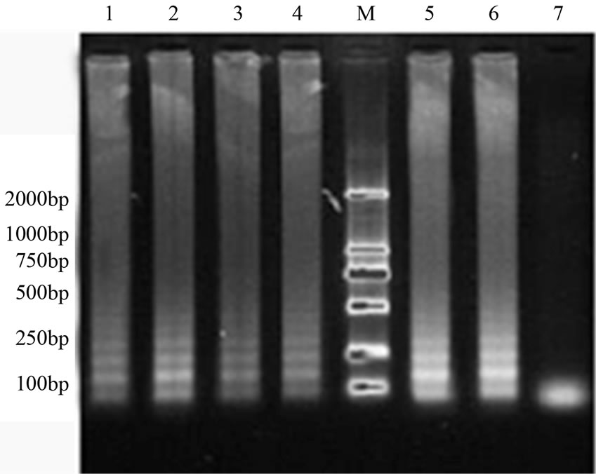
图4表明,MLV酶浓度在6~14 U范围时,均有LAMP反应发生。浓度为4 U时没有LAMP反应发生。最终确定MLV酶的最佳浓度为10 U。
3.1.5. 反应温度优化结果
反应温度在60℃~66℃范围内时,均有LAMP反应发生。当温度在65℃时条带最亮,因此,最终确定最佳反应温度为65℃(图5)。
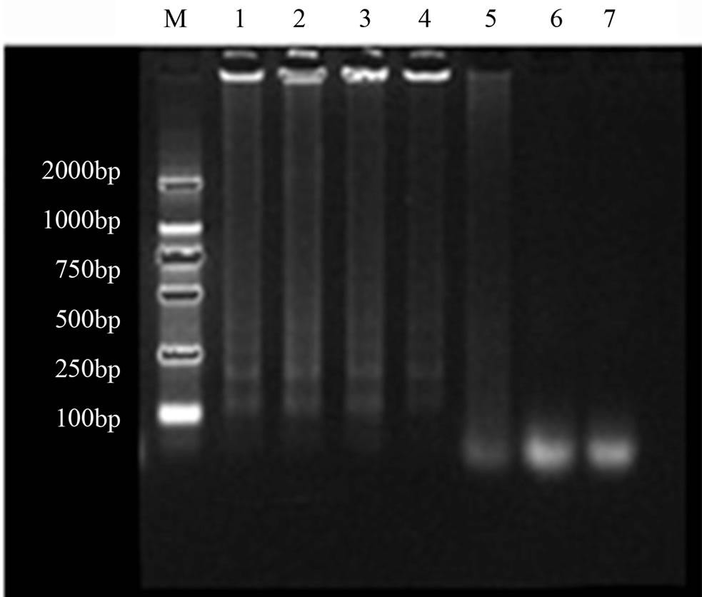
1. 14 U; 2. 12 U; 3. 10 U; 4. 8 U; 5. 6 U; 6. 4 U; 7.negative control; M. marker
Figure 4. The optimized results of MLV enzyme concentration in LAMP reaction
图4. LAMP反应MLV酶浓度优化结果
1. 60℃; 2. 61℃; 3. 62℃; 4. 63℃; 5. 64℃; 6. 65℃; 7. 66℃; 8. negative control; M. marker
Figure 5. The optimized results of reaction temperature in LAMP reaction
图5. LAMP反应温度优化结果
3.1.6. 反应时间优化结果
当反应时间在45 min~120 min范围内,均有LAMP反应发生,时间在45 min时条带较暗,而在60~120 min时间范围时条带亮度较亮且无明显区别。因此最终确定LAMP最佳反应时间为60 min(图6)。
3.2. LAMP扩增产物的电泳检测和染料检测
LAMP扩增产物经琼脂糖凝胶电泳检测,阳性管有特异性的梯状条带,阴性对照则无条带出现(图7)。加入SYBR Green I后,阳性的样本肉眼观察为黄绿色,阴性则观察不到颜色或者为淡橙色(图8)。
1. 45 min; 2. 60 min; 3. 75 min; 4. 90 min; 5. 105 min; 6. 120 min; 7. negative control; M. marker
Figure 6. The optimized results of reaction time in LAMP reaction
图6. LAMP反应时间优化结果
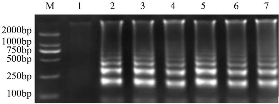
1. negative control; 2-7. HBoV positive sample; M. marker
Figure 7. Analysis of LAMP reaction production using agarose gel electrophoresis
图7. LAMP反应产物的琼脂糖凝胶分析
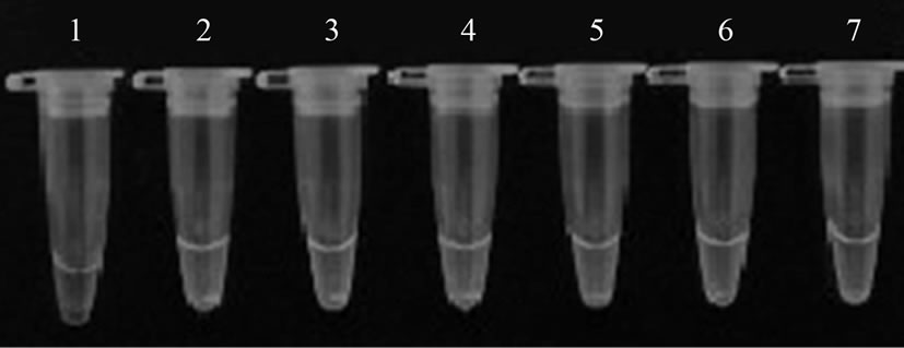
1. negative control; 2-7. HBoV positive sample; M. marker
Figure 8. The results of LAMP amplification (SYBR Green 1 staining)
图8. LAMP扩增结果(SYBR Green 1染色)
3.3. LAMP反应的灵敏性检测
目的条带回收后的产物浓度进行10倍递进稀释得到1.0 × 10⁹~1.0 × 10⁻¹ copies/μL共11个稀释度,LAMP扩增后用SYBR Green I进行检测,当模板浓度在1.0 × 10⁹~1.0 × 100 copies/μL之间显绿色,当模板含量在1.0 × 10−1 copies/μL时显淡橙色,阴性对照也是如此(图9)。表明所建立的LAMP检测方法有很高的检测灵敏性,检测最低可达到1拷贝数。而实时荧光定量PCR检测方法最低检测浓度为100 copies/μL,由此可见LAMP检测方法相对于实时荧光定量PCR,不仅反应时间更快,而且对模板检测也更加灵敏。
3.4. LAMP反应的特异性检测
用所建立的方法对FluA、PIV、RSV和AdV检测结果均为阴性(图10)。表明所建立的LAMP方法具有良好的特异性。
3.5. 实时荧光定量PCR和LAMP检测临床样本
对2012年5~7月间武汉市儿童医院检测儿童的60份咽试子样本,同时用实时荧光定量PCR和LAMP方法进行检测,都检测出8份HBoV阳性样本,二种检测方法的检测结果一致。
4. 讨论
自1985年发明聚合酶链式反应方法[11]已经广泛应用于生物学检测,由高温变性、低温退火以及延伸等几个步骤,使目的DNA迅速扩增。直到2000年,日本科学家Notmoi等[4]发明了环介导等温扩增方法(LAMP),在Bst链式聚合酶的作用下快速完成对靶序列的信号放大。该反应中对引物的设计具有特异性,因此检测方法的特异较高,整个检测过程在65℃等温条件下只需1 h就可完成。
对LAMP扩增产物进行检测时,反应体系中会出现肉眼可视的扩增反应副产物即白色焦磷酸镁沉淀,但由于对Mg2+和引物浓度进行了优化,实验并没有大量的白色沉淀。只有在体系中加入SYBR Green I染料再观察LAMP扩增产物,结果就较为明显了。
在对临床儿童咽试子检测,同时用了实时荧光定量PCR和LAMP检测方法,比较发现,两种检测方法的检出率相同。但进行灵敏度检测时,发现LAMP
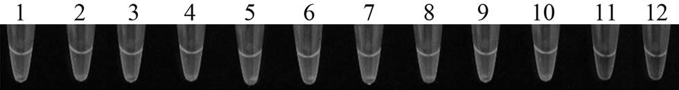
1. 1.0 × 10⁹ copies/Μl; 2. 1.0 × 10⁸ copies/μL; 3. 1.0 × 10⁷ copies/μL; 4. 1.0 × 10⁶ copies/μL; 5. 1.0 × 10⁵ copies/μL; 6. 1.0 × 10⁴ copies/μL; 7. 1.0 × 10³ copies/μL; 8. 1.0 × 10² copies/μL; 9. 1.0 × 101 copies/μL; 10. 1.0 × 100 copies/μL; 11. 1.0 × 10⁻¹ copies/μL; 12. negative control
Figure 9. The sensitivity of LAMP reaction (SYBR Green 1 staining)
图9. LAMP反应的灵敏性检测
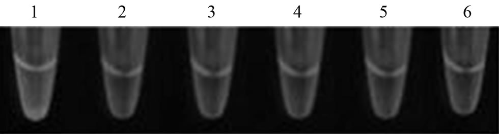
1. HBoV; 2. ddH2O; 3. FluA; 4. PIV; 5. RSV; 6. AdV
Figure 10. The specificity of LAMP reaction(SYBR Green 1 staining)
图10. LAMP反应的特异性检测
检测方法要比PCR检测方法灵敏度高很多[12]。使用LAMP方法检测病毒核酸有很好的便捷性、特异性以及灵敏度。基于操作性、检测时间和对设备要求等方面,LAMP检测方法都优于PCR检测方法,更适合在基层医疗检测中推广。
5. 致谢
感谢国家科技重大专项“湖北及周边省传染病病原谱流行规律研究”(2009ZX10004207; 2012ZX10004 207)的支持。
参考文献 (References)
[1] T. Allander, M. T. Tammi, M. Eriksson, et al. Cloning of a human parvovirus by molecular screening of respiratory tract samples. Proceedings of the National Academy of Sciences of the United States of America, 2005, 102(36): 12891-12896.
[2] X. Ma, R. Endo, N. Ishiguro, et al. Detection of human bocavirus in Japanese children with lower respiratory tract infections. Journal of Clinical Microbiology, 2006, 44(3): 1132-1134.
[3] J. Lindner, S. Modrow. Human bocavirus—A novel parvovirus to infect human. Intervirology, 2008, 51(2): 116-122.
[4] T. Notomi, H. Okayama, H. Masubuchi, et al. Loop-mediated isothermal amplification of DNA. Nucleic Acids Research, 2000, 28(12): E63.
[5] L. L. Poon, C. S. Leung, K. H. Chan, et al. Detection of human influenza A viruses by loop-mediated isothermal amplification. Journal of Clinical Microbiology, 2005, 43(1): 427-430.
[6] 常小斌, 刘洵, 程天印等. 环介导等温扩增技术及其应用研究进展[J]. 畜牧兽医科技信息, 2006, 12: 12-14.
[7] Y. Deng, X. Gu, X. Zhao, et al. High viral load of human bocavirus correlates with duration of wheezing in children with severe lower respiratory tract infection. PLoS One, 2012, 7(3): e34353.
[8] 张坤, 黄伟, 李刚. H5N1禽流感病毒环介导等温扩增快速检测方法的建立[J]. 生物技术通讯, 2009, 20(2): 217-220.
[9] T. Okafuji, N. Yoshida, M. Fujino, et al. Rapid diagnostic method for detection of mumps virus genome by loop-mediated isothermal amplification. Journal of Clinical Microbiology, 2005, 43(4): 1625-1631.
[10] M. Ito, M. Watanabe, N. Nakagawa, et al. Rapid detection and typing of influenza A and B by loop-mediated isothermal amplification: Comparison with immunochromatography and virus isolation. Journal of Virological Methods, 2006, 135(2): 272- 275.
[11] 李坤, 史伟峰, 石旦等. 逆转录–环介导等温扩增快速检测EV71病毒[J]. 分子诊断与治疗杂志, 2012, 4(1): 26-29.
[12] N. Mori, Y. Motegi, Y. Shimamura, et al. Development of a new method for diagnosis of rubella virus infection by reverse transcription-loop-mediated isothermal amplification. Journal of Clinical Microbiology, 2006, 44(9): 3268-3273.
NOTES
*通讯作者。