International Journal of Psychiatry and Neurology
Vol.04 No.04(2015), Article ID:16332,6
pages
10.12677/IJPN.2015.44005
Prognostic Value of Plasma Brain Natriuretic Peptide Levels in Patients with Acute Intracerebral Hemorrhage
Daoming Wei1, Xuehong Zhu1, Lili Gao2, Ruiling Yang2, Jushan Lin2, Yuansheng Liao2, Shiju Li2, Chenghan Wu2*
*通讯作者。
1Department of Endocrinology, 85 Hospital of People’s Liberation Army, Shanghai
2Department of Neurology, The Second Affiliated Clinical College of Fujian University of Traditional Chinese Medicine, Fuzhou Fujian
Received: Oct. 26th, 2015; accepted: Nov. 10th, 2015; published: Nov. 13th, 2015
Copyright © 2015 by authors and Hans Publishers Inc.
This work is licensed under the Creative Commons Attribution International License (CC BY).
http://creativecommons.org/licenses/by/4.0/


ABSTRACT
Objective: To study brain natriuretic peptide (BNP) levels and prognosis in patients with cerebral hemorrhage. Methods: Retrospective analysis of 109 cases with cerebral hemorrhage from December 2010 to December 2014 for the treatment was divided into 2 groups, according to the outcome of a good outcome group and poor outcome groups. The enzyme-linked immunosorbent assay (ELISA) was used for determination of plasma BNP. Result: There was a close relationship between patient outcomes and a previous history of stroke, smoking history, hematoma volume, Glasgow Coma Scale score, National Research Health Stroke Scale score, rupture of hematoma into the ventricle, plasma brain natriuretic peptide level. (X2 = 3.678, 7.096, t = 2.962, 2.361, 3.806, X2 = 4.687, t = 4.962, P < 0.05); Logistic regression analysis revealed a correlation between the prognosis of brain natriuretic peptide levels in patients with acute cerebral hemorrhage (P = 0.032, OR = 1.789, 95% CI 1.052 - 3.043). Conclusions: Plasma brain natriuretic peptide level is an independent risk factor for patients with cerebral hemorrhage and may be a useful prognostic factor for these patients.
Keywords:Cerebral Hemorrhage, Brain Natriuretic Peptide, Prognosis

血浆脑钠肽水平对急性脑出血患者转归的预测价值
韦道明1,朱雪红1,高丽丽2,杨瑞玲2,林菊珊2,廖远生2,李世举2,吴成翰2*
1中国人民解放军八五医院神经内分泌科单位,上海
2福建中医院大学附属第二人民医院神经内科,福建 福州

收稿日期:2015年10月26日;录用日期:2015年11月10日;发布日期:2015年11月13日

摘 要
目的:探讨血浆脑钠肽(BNP)水平与脑出血患者预后的关系。方法:回顾性分析2010年12月~2013年12月治疗的109例脑出血患者,根据转归分为转归良好组和转归不良组。采用酶联免疫吸附法(ELISA)测定血浆BNP。结果:患者卒中病史、吸烟史、血肿体积、格拉斯哥昏迷评分、美国国立卫生研究量卒中量表评分、血肿是否破入脑室及血浆脑钠肽水平均与转归结果有密切关系(X2 = 3.678, 7.096, t = 2.962, 2.361, 3.806, X2 = 4.687, t = 4.962, P < 0.05); Logistic回归分析发现血浆脑钠肽水平与急性脑出血患者的预后相关(P = 0.032, OR = 1.789, 95% CI 1.052~3.043)。结论:血浆BNP是脑出血患者转归的危险因素之一,对患者预后的判断有一定的价值。
关键词 :脑出血,脑钠肽,预后

1. 引言
脑出血是常见病、多发病,病情发展迅速,预后较差,后遗症较多,对患者生存质量造成了严重影响。高血压和动脉硬化是脑出血的主要因素。脑出血后除了占位效应损伤外,还与早期的继发性损伤有关,如:血浆蛋白渗出和血凝块回缩[1] [2] ,基质金属蛋白酶及凝血酶的改变 [3] [4] 以及血红蛋白的释放及其降解产物如血红素、铁离子、铁蛋白等产生的损害作用外 [5] - [11] ,还与细胞凋亡有关 [12] - [16] ,影响其预后的因素决定于:出血部位、出血量、年龄及感染的严重程度 [17] ,有关使用实验室检查的结果来预测预后的研究鲜有报道 [18] 。B型脑钠肽(brain natriuriritie Peptide, BNP)是脑钠肽家族中研究较多的生物活性物质,具有强大的排钠、利尿、舒张血管和抑制肾素–醛固酮系统等作用 [19] 。BNP在体内除存在于周围组织中,也存在于中枢神经系统内,主要分布在心、肺、脑、脊髓和垂体以及血浆和脑脊液中。其中心脏的BNP含量最高,其次是脑。在脑中的含量以延脑、纹壮体及下丘脑等处最多 [20] 。研究表明 [21] ,BNP参与了高血压与颅高压的病理生理过程,且在脑出血后迅速升高。颅脑CT检查对脑出血和出血量的检出有决定性的作用,但出血量与预后不一定具有平行关系。因此,寻找一种能够对患者病情及预后作出有效评估的指标具有重要临床意义。以往研究表明BNP与脑梗死后的脑水肿有关,是反映预后的良好指标 [22] 。本研究采用回顾性研究的方法,通过对109例脑出血患者血浆BNP水平及其与病情及预后关系进行了观察,现将结果报告如下。
2. 资料与方法
2.1. 脑出血病人
总共109例,来自2010年12月~2014年12月分别在中国人民解放军八五医院神经内分泌科和福建中医院大学附属第二人民医院神经内科住院治疗的脑出血患者,纳入标准:1) 全部病例均经头部CT或MRI证实,符合1996年全国第四届脑血管病学术会标准。2) 患者均为临床首次脑出血,既往无影响神经系统的疾病。3) 发病24 h内就诊者。4) 既往无严重心、肺、肝、肾等重要脏器疾病的患者。其中男67例,女42例,年龄38~67岁,平均年龄(51.5 ± 4.6)岁;入院后50例进行了开颅血肿消除术或微创血肿清除术,59例进行了内科保守治疗。其中转归良好组72例,转归不良组37例。
2.2. 资料收集
所有入组的病例均在入院时进行格拉斯哥昏迷量表评分(GCS)、美国国立卫生研究量卒中量表评分(NIHSS),根据多田氏公式(π/6 × 长 × 宽 × 层数)计算出血量,并记录其血肿部位及血肿是否破入脑室、既往是否合并高血压病、糖尿病、高脂血症、卒中或TIA、脑出血、缺血性心脏病、吸烟、饮酒、发病前是否使用抗血小板药或抗凝药。
2.3. 血浆BNP检测
入选脑出血患者均于入院后24 h内采集肘静脉血2 ml,置入含有乙二胺四乙酸(EDTA)的试管中抗凝,采用1500 r/min离心5 min分离血浆,置入−30℃冰箱保存统一待测,BNP血浆浓度采用酶联免疫吸附试验(ELISA)检测,试剂盒采用上海拜力生物科技有限公司提供的人BNP ELISA试剂盒。
2.4. 疗效评定
所有患者均于发病后3个月通过改良Rankin量表(modified Rankin Scale, mRS)进行疗效评定,mRS评分 <= 2分为转归良好,>2分为转归不良。
2.5. 统计学方法
采用SPSS 11.5统计软件对所有实验数据进行整理与分析,计数资料的比较采用卡方检验,计量资料数据用(x ± s)表示,两样本均数比较采用t检验。P < 0.05定义为有统计学意义,分析影响因素时采用Logistic回归方法(以P < 0.05为有统计学意义)。
3. 结果
本研究共选取符合条件的109例脑出血患者,所有患者均完成随访,无脱落患者。按转归分为转归良好组和转归不良组,其中转归良好组72例(66%),转归不良组37例(34%)。两组患者在年龄、性别、既往合并高血压病史、既往合并高脂血症、糖尿病、脑出血史、缺血性心脏病、饮酒、入院收缩压、入院舒张压、入院空腹血糖、发病前是否使用抗凝药物、治疗方案、血肿部位均无统计学差异,P > 0.05 (见表1)。而在卒中病史、吸烟史、入院血肿体积、入院格拉斯哥昏迷评分、入院美国国立卫生研究量卒中量表评分、血肿是否破入脑室方面进行比较,两组间具有显著性差异(X2 = 3.678, 7.096, t = 2.962, 2.361, 3.806, X2 = 4.687, P < 0.05)。两组患者入院血浆脑钠肽的水平,转归良好组为(826.3 ± 163.1) pg/ml,转归不良组为(1572.4 ± 271.5) pg/ml,两组具有显著差异(t = 4.962, P < 0.05),采用Logistic回归分析发现血浆脑钠肽水平与急性脑出血患者的预后相关(P = 0.032, OR = 1.789, 95% CI 1.052~3.043)。表明患者的高血压病史、既往卒中病史、吸烟史、血肿体积、格拉斯哥昏迷评分、国立卫生研究量卒中量表评分、血肿是否破入脑室、血浆脑钠肽均与转归是否良好具有密切的关系。
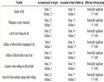
Table 1. Demographic and clinical data of the study population
表1. 研究对象的人口统计学和临床资料
4. 讨论
脑出血是一种常见的脑血管疾病,死亡率较高,诸多因素与其预后有着密切的联系。及时判断患者预后对临床治疗有着重要的意义。脑钠肽是一种生物活性肽,由32个氨基酸组成,其主要的作用有排钠、利尿及其抑制肾素–血管紧张素–醛固酮系统等,在许多疾病的生理病理中扮演了重要的角色 [19] 。BNP主要来源于心室,各种原因造成的容量负荷增加和心室壁张力都会造成其生成和分泌增加。BNP的含量在脑内仅次于心肌,在各种脑血管疾病导致的脑水肿、颅内压增高时,均可增加BNP的分泌,提示在颅内高压的病理生理过程中均有BNP的参与 [21] 。Berendes [23] 、Fukui [24] 、Espiner等 [25] 均报道患者发生蛛网膜下腔出血后血浆BNP水平均有明显增高,并认为脑血管痉挛、低钠血症均与血清脑钠肽增高有一定相关性。McGirt MJ等 [26] 报道血清脑钠肽与蛛网膜下腔出血后低钠血症和延迟性神经损伤的相关性,结果血清脑钠肽增高与低钠血症无关,而延迟缺血性神经损伤发生后头24小时血清脑钠肽显著升高,可以预测2周后的GCS评分,这类病人由于严重的脑水肿更容易影响下丘脑一垂体系统导致神经介质的分泌异常。也有研究表明,BNP在脑梗死后导致的脑水肿患者中明显增加,是反映患者预后的指标 [27] 。
本研究显示,脑钠肽水平与脑出血患者的转归有密切的关系,是脑出血预后评定的独立危险因素,原因可能是脑出血发生后,许多活性物质(如红细胞降解产物、凝血酶 [4] 、补体、五羟色氨等)进入到血液中,脑血管和组织受到持续刺激,同时由于血肿挤压和牵拉周围的组织和血管,使脑组织发生继发性缺血缺氧,刺激脑钠肽的产生。与Cameron V. A.等 [28] 报道一致,BNP在急性脑血管意外患者中可异常增高,其增高程度与疾病严重程度平行,并与预后相关,可望作为急性脑血管意外的病情评价和转归预测的良好指标;James M. L.等 [29] 的研究也发现脑出血患者BNP的水平能准确反映他们的神经功能预后,Modrego P. J.等[30] 推断脑出血患者BNP的水平可反映亚急性期脑水肿状况。本研究结果显示,血浆BNP可作为脑出血后预后评估的独立危险因素,与国外学者研究结果相符,有望为脑出血预后的评估建立新的客观证据,进一步指导脑出血患者的治疗,改善预后。
基金项目
本课题得到以下基金资助:国家自然基金81403255,国家自然基金81503431,福建省教育厅A类科技重点课题JA11141,福建省卫生厅医学创新课题2011-cx-29,福建省自然科学基金2011j01196,福建省卫生厅中医药临床基地课题zlcx03,福建省引智项目基金20153500016。福建省卫生和计划生育委员会青年科研课题2015-1-85。
文章引用
韦道明,朱雪红,高丽丽,杨瑞玲,林菊珊,廖远生,李世举,吴成翰. 血浆脑钠肽水平对急性脑出血患者转归的预测价值
Prognostic Value of Plasma Brain Natriuretic Peptide Levels in Patients with Acute Intracerebral Hemorrhage[J]. 国际神经精神科学杂志, 2015, 04(04): 28-33. http://dx.doi.org/10.12677/IJPN.2015.44005
参考文献 (References)
- 1. Wagner, K.R. and Dwyer, B.E. (2004) Hematoma Removal, Heme, and Heme Oxygenase Following Hemorrhagic Stroke. Annals of the New York Academy of Sciences, 1012, 237-251. http://dx.doi.org/10.1196/annals.1306.020
- 2. Xi, G.H., Wagner, K.R., Keep, R.F., et al. (1998) Role of blood Clot Formation on Early Edema Development after Experimental Intracerebral Hemorrhage. Stroke, 29, 2580-2586. http://dx.doi.org/10.1161/01.STR.29.12.2580
- 3. Wu, C.H., Huang, F.Y., Wang, K.Y., et al. (2008) Expression of Matrix Metalloproteinase MMP-9 in the Plasma and Hematoma Fluid of Intracerebral Hemorrhage Patients. National Medical Journal of China, 88, 174-176.
- 4. Wu, C.H., Yang, R.L., Huang, S.Y., et al. (2011) Analysis of Thrombin-Antithrombin Complex Contents in Plasma and Hematoma Fluid of Hypertensive Intracerebral Hemorrhage Patients after Clot Removal. European Journal of Neurology, 18, 1060-1066. http://dx.doi.org/10.1111/j.1468-1331.2010.03336.x
- 5. Xi, G., Hua, Y., Bhasin, R.R., Ennis, S.R., Keep, R.F. and Hoff, J.T. (2011) Mechanisms of Edema Formation after Intracerebral Hemorrhage: Effects of Extravasated Red Blood Cells on Blood Flow and Blood-Brain Barrier Integrity. Stroke, 32, 2932-2938. http://dx.doi.org/10.1161/hs1201.099820
- 6. Lara, F.A., Kahn, S.A., da Fonseca, A.C., et al. (2009) On the Fate of Extracellular Hemoglobin and Heme in Brain. Journal of Cerebral Blood Flow & Metabolism, 29, 1109-1120. http://dx.doi.org/10.1038/jcbfm.2009.34
- 7. Song, S., Hua, Y., Keep, R.F., et al. (2007) A New Hippocampal Model for Examining Intracerebral Hemorrhage-Re- lated Neuronal Death: Effects of Deferoxamine on Hemoglobin-Induced Neuronal Death. Stroke, 38, 2861-2863. http://dx.doi.org/10.1161/STROKEAHA.107.488015
- 8. Regan, R.F., Chen, M., Li, Z., et al. (2008) Neurons Lacking Iron Regulatory Protein-2 Are Highly Resistant to the Toxicity of Hemoglobin. Neurobiology of Disease, 31, 242-249. http://dx.doi.org/10.1016/j.nbd.2008.04.008
- 9. Mehdiratta, M., Kumar, S., Hackney, D., et al. (2008) Association between Serum Ferritin Level and Perihematoma Edema Volume in Patients with Spontaneous Intracerebral Hemorrhage. Stroke, 39, 1165-1170. http://dx.doi.org/10.1161/STROKEAHA.107.501213
- 10. Selim, M. (2009) Deferoxamine Mesylate: A New Hope for Intracerebral Hemorrhage: From Bench to Clinical Trials. Stroke, 40, 90-91. http://dx.doi.org/10.1161/STROKEAHA.108.533125
- 11. Qing, W.G., Dong, Y.Q., Ping, T.Q., et al. (2009) Brain Edema after Intracerebral Hemorrhage in Rats: The Role of Iron Overload and Aquaporin 4. Journal of Neurosurgery, 110, 462-468. http://dx.doi.org/10.3171/2008.4.JNS17512
- 12. Wu, C.H., Ding, X.Y., Ye, X.B., et al. (2009) Neural Apoptosis of Around Hematoma and Some Apoptosis-Gene in Intracerebral Hemorrhage Patients. Journal of Internal Medicine of Taiwan, 20, 440-446
- 13. Felberg, R.A., Grotta, J.C., Shirzadi, A.L., et al. (2002) Cell Death in Experimental Intracerebral Hemorrhage: The “Black Hole” Model of Hemorrhagic Damage. Annals of Neurology, 51, 517-524. http://dx.doi.org/10.1002/ana.10160
- 14. Qureshi, A.I., Suri, M.F., Ostrow, P.T., et al. (2003) Apoptosis as a Form of Cell Death in Intracerebral Hemorrhage. Neurosurgery, 52, 1041-1047; Discussion 1047-1048. http://dx.doi.org/10.1227/01.neu.0000057694.96978.bc
- 15. Xue, M. and Del Bigio, M.R. (2000) Intracortical Hemorrhage Injury in Rats: Relationship between Blood Fractions and Brain Cell Death. Stroke, 31, 1721-1727. http://dx.doi.org/10.1161/01.STR.31.7.1721
- 16. Matsushita, K., Meng, W., Wang, X., et al. (2000) Evidence for Apoptosis after Intercerebral Hemorrhage in Rat Striatum. Journal of Cerebral Blood Flow & Metabolism, 20, 396-404. http://dx.doi.org/10.1097/00004647-200002000-00022
- 17. Diedler, J., Sykora, M., Hahn, P., et al. (2009) C-Reactive-Protein Levels Associated with Infection Predict Short- and Long-Term Outcome after Supratentorial Intracerebral Hemorrhage. Cerebrovascular Diseases, 27, 272-279. http://dx.doi.org/10.1159/000199465
- 18. Löppönen, P., Qian, C., Tetri, S., et al. (2014) Predictive Value of C-Reactive Protein for the Outcome after Primary Intracerebral Hemorrhage. Journal of Neurosurgery, 29, 1-6. http://dx.doi.org/10.3171/2014.7.jns132678
- 19. Chusho, H., Tamura, N., Ogawa, Y., et al. (2001) Dwarfism and Early Death in Mice Lacking C-Type Natriuretic Peptide. Proceedings of the National Academy of Sciences of the United States of America, 98, 4016-4021. http://dx.doi.org/10.1073/pnas.071389098
- 20. Lang, C.C., Choy, A.M. and Struthers, A.D. (1992) Atrial and Brain Natriuretic Peptides: A Dual Natriuretic Peptide System Potentially Involved in Circulatory Homeostasis. Clinical Science (London), 83, 519-527. http://dx.doi.org/10.1042/cs0830519
- 21. Stewart, D., Waxman, K., Brown, C.A., et al. (2007) B2 Type Natriuretic Peptide Levels May Be Elevated in the Critically Injured Trauma Patient without Congestive Heart Failure. Journal of Trauma, 63, 747-750. http://dx.doi.org/10.1097/01.ta.0000240458.46050.38
- 22. Tokudome, T., Kishimoto, I., Yamahara, K., et al. (2009) Impaired Recovery of Blood Flow after Hind-Limb Ischemia in Mice Lacking Guanylyl Cyclase-A, a Receptor for Atrial and Brain Natriuretic Peptide. Arteriosclerosis, Thrombosis, and Vascular Biology, 29, 1516-1521. http://dx.doi.org/10.1161/ATVBAHA.109.187526
- 23. Berendes, E., Van-Aken, H., Raufhake, C., et al. (2010) In Diferential Secretion of Atrial and Brain Natriuretic Pepfide in Crifitally Ill Patients. Anesthesia & Analgesia, 93, 676-682. http://dx.doi.org/10.1097/00000539-200109000-00029
- 24. Fukui, K., Lnamura, T., Nakamizo, A., et al. (2000) Relationship between Cardiac Natriuretic Peptide (ANP/BNP) and Fluid Intake in Patients with Subarachnoid Hemorrhage. No To Shinkei Brain and Nerve, 52, 1019-1023.
- 25. Espiner, E.A., Leikis, R., Fetch, R.D., et al. (2009) The Neu-ro-Cardioendocrine Response to Acute Subaraehnoid Haemorhage. Clinical Endocrinology (Oxford), 56, 629-635. http://dx.doi.org/10.1046/j.1365-2265.2002.01285.x
- 26. McGirt, M.J., Blessing, R., Nimjee, S.M., et al. (2004) Correlation of Serum Brain Natriuretic Peptide with Hyponatremia and Delayed Ischemic Neurological Deficits after Subarachuoid Hemorrhage. Neurosurgery, 54, 1369-1373; Discussion 1373-1374. http://dx.doi.org/10.1227/01.NEU.0000125016.37332.50
- 27. Isotani, E., Suzuki, R., Tomita, K., et al. (1994) Alterations in Plasma Concentrations of Natriuretic Peptides and Antidiuretic Hormone after Subarachnoid Hemorrhage. Stroke, 25, 2198-2203. http://dx.doi.org/10.1161/01.STR.25.11.2198
- 28. Cameron, V.A. and Richards, A.M. (2002) Natriuretic Peptide System in Fetal Heart and Circulation. Journal of Hypertension, 20, 801-803. http://dx.doi.org/10.1097/00004872-200205000-00003
- 29. James, M.L., Blessing, R., Phillips Bute, B.G., et al. (2009) S100B and Brain Natriuretic Peptide Predict Functional Neurological Outcome after Intracerebral Haemorrhage. Biomarkers, 14, 388-394. http://dx.doi.org/10.1080/13547500903015784
- 30. Modrego, P.J., Boned, B., Berlznga, J.J., et al. (2008) Plasmatic B-Type Natriuretic Peptide and C-Reactive Protein in Hyperacute Stroke As Markers of CT-Evidence of Brain Edema. International Journal of Medical Sciences, 5, 18-23. http://dx.doi.org/10.7150/ijms.5.18