Open Journal of Natural Science
Vol.
09
No.
02
(
2021
), Article ID:
40809
,
9
pages
10.12677/OJNS.2021.92023
基于碳量子点内滤作用测定水样中对硝基苯酚
邵悦馨,张云龙,王鑫,王林慧,王翠翠,王菁星,向国强*
河南工业大学化学化工学院,河南 郑州

收稿日期:2021年1月6日;录用日期:2021年2月26日;发布日期:2021年3月5日

摘要
以蚂蚱粉末为碳源,利用硝酸与乙二胺酸碱反应的反应热实现快速、简便制备碳量子点(CDs)。所制备的CDs的最佳激发波长为390 nm,发射峰为470 nm。由于对硝基苯酚(p-NP)在400 nm处具有特征吸收,与CDs的荧光激发峰具有良好的光谱重叠,使p-NP对CDs具有明显的内滤作用,且其它硝基苯化合物的吸收峰几乎不与CDs荧光发射峰重叠,因此不产生荧光内滤作用。基于该荧光内滤作用,建立了p-NP的高灵敏度,高选择性荧光分析新方法。该分析方法检出限(3σ)为0.13 μM,相对标准偏差(RSD)为4.09% (c = 10 μM, n = 11),线性范围是0.5~80 μM。该方法已成功应用于环境水样中痕量p-NP的检测。
关键词
碳量子点,内滤作用,环境水样,对硝基苯酚

Determination of P-Nitrophenol in Water Samples Based on Carbon Dots by Inner Filter Effect
Yuexin Shao, Yunlong Zhang, Xin Wang, Linhui Wang, Cuicui Wang, Jingxing Wang, Guoqiang Xiang*
School of Chemistry and Chemical Engineering, Henan University of Technology, Zhengzhou Henan

Received: Jan. 6th, 2021; accepted: Feb. 26th, 2021; published: Mar. 5th, 2021

ABSTRACT
Carbon Dots (CDs) were prepared using locust powder as carbon source through the exothermic reaction between nitric acid and ethylenediamine. The optimal excitation wavelength and emission peak of CDs prepared were 390 nm and 470 nm respectively. Due to the characteristic absorption of p-Nitrophenol (p-NP) at 400 nm, which has a good spectral overlap with the fluorescence excitation peak of CDs, then P-NP has an obvious Inner Filter Effect (IFE) on the fluorescence emission of CDs. Moreover, the absorption peak of other nitrobenzene compounds hardly overlaps with the fluorescence excitation and emission peak of CDs, so no IFE is generated. A new analytical method for p-NP was established based on IFE with high sensitivity and selectivity. The detection limit (3σ) of the method for p-NP was 0.13 μM, and Relative Standard Deviation (RSD) was 4.09% (c = 10 μM, n = 11), and the linear range was 0.5~80 μM. The method has been successfully applied to the determination of trace p-NP in environmental water samples.
Keywords:Carbon Dots, Inner Filter Effect, Environmental Water Samples, P-Nitrophenol

Copyright © 2021 by author(s) and Hans Publishers Inc.
This work is licensed under the Creative Commons Attribution International License (CC BY 4.0).
http://creativecommons.org/licenses/by/4.0/


1. 引言
对硝基苯酚(p-NP)是一种广泛应用于染料、农药、炸药、杀菌剂、医药等领域的重要的硝基芳香化合物 [1] [2]。由于p-NP的生物降解性差、稳定性好、具有生物累积性,从污染的水体和土壤中去除p-NP是非常困难的。p-NP可能导致严重的健康问题,一旦进入血液可以将血红蛋白转化为高铁血红蛋白,从而导致肝损伤、贫血、呼吸困难,还有其他相关症状如头痛、肾脏和中枢神经系统损伤 [3] [4],更严重的还会导致基因突变和致癌 [5]。美国环境保护署(EPA)将p-NP列为“优先污染物”,限制了其在水中最大浓度不超过10 ng∙L−1 [6]。
目前,监测环境中p-NP的分析方法主要有,毛细管电泳法 [7],色谱分析方法 [8],电化学分析法 [9],但是这些分析方法有一些缺点,比如电极不稳定,需要昂贵的大型仪器,需要训练有素的操作人员以及复杂的前处理过程等 [10]。与上述分析方法相比,荧光传感分析由于具有高选择性和高灵敏度、分析速度快、操作简便、分析成本低等优点而备受关注 [11] [12] [13]。许多荧光纳米材料,如CDS [14] [15] [16] [17]、ZnOQDS [18] 和CdTeQDS [19] [20],都被用于构建p-NP的荧光传感分析方法。
CDs是一类新的荧光纳米材料,与发光量子点(QDs)和有机荧光染料相比,由于其优异的化学稳定性,生物相容性,低毒性和可调光致发光的特性而受到广泛关注 [21] [22],在荧光传感分析和生物成像分析中获得了广泛应用 [23] [24] [25]。已有基于CDs的p-NP荧光传感分析报道,然而这些报道中CDs大多采用高耗能、多费时的水热制备方法,且分离纯化较为复杂。
本文以蚂蚱粉末为碳源,利用硝酸与乙二胺的酸碱反应实现热裂解制备CDs,由于CDs的激发谱带(390 nm)与p-NP在400 nm处的吸收峰有良好的光谱重叠,这使得p-NP对CDs的荧光具有强烈的内滤效应(IFE)。基于IFE建立了p-NP分析的新方法,成功将该方法应用于水样中p-NP的检测。
2. 实验部分
2.1. 试剂和仪器
碳量子点(CDs)的透射电子显微镜(TEM)图像通过JEM-2011透射电子显微镜获得,使用荧光光谱仪(RF-6000,岛津,日本)收集激发和发射光谱,使用FL-TCSPC荧光光谱仪(Horiba JobinYvon,法国)测量CDs的荧光寿命,在WQF-510傅里叶变换红外(FT-IR)光谱仪(北京北分瑞利分析仪器(集团)有限公司,中国北京)上记录了CDs红外吸收光谱,CDs的UV-Vis光谱使用UV-紫外可见光谱仪(UV2600,岛津,日本)获得。
硝基苯酚(p-NP)购自上海阿拉丁生化科技股份有限公司。六水合氯化铁(FeCl3·6H2O)购于天津市科密欧化学试剂有限公司。乙二胺(EDA),金属盐(除FeCl3·6H2O),果糖(D-Fru),甘露糖(D-Man),葡萄糖(Glu),苯丙氨酸(Phe),甲硫氨酸(Met),组氨酸(His),乳果糖(Lac)有机化合物,2,4-二氯苯酚(DCP),对氯苯酚(p-CP),硝基苯(NB),对硝基甲苯(p-NT),2,4-二硝基苯酚(DNP)硝基芳香族化合物购自国药控股化学试剂有限公司(中国上海)。蚂蚱粉末通过家用粉碎机处理干燥的蚂蚱(河南工业大学粮油食品学院提供)得到。超纯水由Milli-Q净化设备(Millipore,美国)(18 MΩ cm−1)提供,整个实验过程中Phosphate Buffer (P-B)缓冲溶液用于整个实验酸度的控制。其它化学试剂均为分析纯,使用前未进行任何纯化处理。
2.2. CDs的制备
利用HNO3与乙二胺之间的酸碱反应放热,实现碳源的热裂解,从而达到制备CDs的目的。具体方法如下:称取0.5 g干燥的蚂蚱粉末于烧杯(100 mL)中,然后向烧杯中加入5 mL乙二胺震荡混合均匀,此时溶液颜色逐渐加深至深棕色,待混合均匀后,再向烧杯中缓慢滴加浓硝酸(5 mL),并振荡溶液。随着酸的加入,溶液在几秒内放出大量的热,沸腾并伴有烟雾产生(于3 min内将HNO3滴加完毕)。将获得的反应液自然冷却至室温,加入超纯水稀释至50 mL,在所得反应液中加入活性碳粉(5 g),磁力搅拌30 min后,将离心分离收集的溶液,经冷冻干燥得到棕黄色CDs粉末。所得CDs粉末溶于一定量超纯水中,制成CDs水溶液(16.0 mg∙mL−1)用于后续实验。
2.3. 实验方法
将0.8 mL CDs溶液(16.0 mg∙mL−1)与一定量的p-NP标准溶液(或样品溶液)分别加入到10 mL比色管中,然后用PB缓冲液(pH 10)定容至所需体积,混合均匀,在390 nm的激发波长下测定存在(I)和不存在(I0)p-NP时的荧光强度(发射峰470 nm),计算两者的lg(I0/I)。
3. 结果与讨论
3.1. CDs的表征
图1(a)是CDs的TEM图,从图上可知CDs都具有典型的球形结构,平均粒径为2.25 nm。利用FT-IR光谱确认了CDs表面的官能团,如图1(b)所示,在769.49 cm−1处有一个吸收峰,它是C=C的拉伸振动峰,在1384.64 cm−1、1766.48 cm−1处的吸收峰分别归因于C-N,C=O的弯曲振动引起的,3016.49 cm−1和3431.83 cm−1处的双宽峰与N-H和O-H伸缩振动有关,以上结果表明已经成功合成CDs。
图1(c)显示了在不同激发波长(300~450 nm)条件下CDs的荧光发射光谱。可以看出CDs的发射光谱表现出明显的激发波长依赖性,在激发波长300~450 nm的范围内,荧光发射波长,随着激发波长的增加而红移。荧光发射强度(Ex 300~390 nm)也随着激发光增加而增加,在390 nm处达到最大发射强度,之后随激发波长进一步增加而减弱。CDs的荧光光谱显示,最佳激发波长为390 nm,最大发射峰位于470 nm。
图1(d)所示,研究了pH对CDs荧光强度的影响。CDs的荧光强度在酸性介质中,具有较强荧光,且随pH增大荧光强度变化不大;CDs的荧光强度在碱性介质中,明显降低,在强碱性条件(pH > 11)下,CDs荧光强度继续极大的降低。
![]() (a)
(a)
![]() (b)
(b)
![]() (c)
(c)
![]() (d)
(d)
Figure 1. (a) Typical TEM of the prepared CDs. (b) FT-IR spectra of the prepared CDs. (c) Emission spectra of CDs at excitation wavelengths from 300~450 nm. (d) Effects of pH on fluorescence intensity of the CDs
图1. (a) CDs的TEM图像,(b) FT-IR光谱,(c) CDs在不同激发波长(300~450 nm)条件下的发射光谱图,(d) 在不同pH条件下CDs的荧光强度
3.2. p-NP的荧光传感分析
3.2.1. CDs对p-NP的传感分析性能
由于p-NP在碱性条件下,在400 nm处有明显的吸收峰,与CDs的激发光谱有较大重叠。实验发现,在pH10条件下,p-NP对CDs荧光具有猝灭作用。
如图2所示,在不同浓度的p-NP条件下,CDs的荧光强度被不同程度的猝灭。随着p-NP浓度的增加,CDs的荧光强度逐渐降低。p-NP浓度(0.5 μM~80 μM)与lg(I0/I)之间呈良好的线性关系,线性方程是lg(I0/I) = 0.0106C + 0.0082,相关系数R2为0.9994,相对标准偏差(RSD)为4.09% (c = 10 μM),根据IUPAC的定义(3σ),该方法的检出限为0.13 μM。表1列出了文献报道的不同荧光探针检测p-NP的分析性能,可以看出本方法具有与其他分析方法相当的线性范围和检出限;不过,与其他文献报道的荧光探针复杂、繁琐、耗时的制备过程相比,本方法所用CDs制备方法简单、快速,制备与分离纯化仅需要40 min。
3.2.2. p-NP传感分析的选择性
为了评估CDs对p-NP的选择性,研究了硝基芳香族化合物、有机化合物和金属离子对CDs荧光强度的影响。如图3(a)和图3(b)所示,浓度为10 μM的硝基芳香族化合物(NB,DCP,p-NT,p-CP,DNP),浓度为100 μM的有机物(Lac,Glu,Met,His,D-Man),及浓度为100 μM的金属离子对存在或不存在p-NP (10 μM)的CDs的荧光几乎没有影响(Hg2+,Cu2+,Fe2+浓度为10 μM时则对其荧光几乎没有影响)。结果表明CDs对p-NP的选择性较高,为检测水样中p-NP提供了实验依据。
![]()
Figure 2. Fluorescence quenching of CDs by p-NP (0.5~80 μM)
图2. p-NP (0.5~80 μM)对CDs的荧光内滤作用(IFE)

Table 1. Comparative data from some studies on fluorescent probes for p-NP
表1. 不同纳米荧光探针检测p-NP的方法及性能
![]()
Figure 3. Interferences of (a) other nitroaromatics, organic compoundsand (b) metal ions in PB buffer
图3. (a) 硝基芳香族化合物,有机化合物及(b) 金属离子对CDs分析体系的影响
3.3. p-NP的传感机理
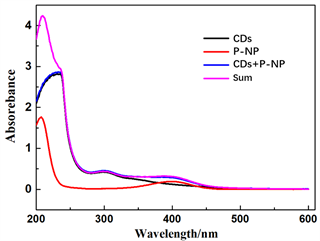
为了推断p-NP对CDs可能的荧光猝灭机理,测定了CDs的荧光发射光谱和p-NP的紫外–可见吸收光谱(图4(a))。从图4(a)可以看出,p-NP的吸收光谱(400 nm处的吸收峰)与CDs的荧光发射光谱之间存在较大的光谱重叠,这可能是由IFE或FRET引起的荧光猝灭机制。为了进一步确定荧光猝灭机理,可用Stern-Volmer方程表示猝灭效率:F0/F = Ksv[Q] + 1 = Kqτ0[Q] + 1,其中F和F0分别是有和没有猝灭剂时的荧光强度,[Q]是猝灭剂浓度,Ksv是Stern-Volmer猝灭常数(M−1),Ksv = Kqτ0,其中Kq是分子猝灭常数,τ0是没有猝灭剂的荧光寿命。对CDs-p-NP体系,Ksv = 0.04962 μM−1和τ0 = 2.90 ns,计算得Kq = 1.71 × 10¹³ M−1 s−1,高于最大扩散控制的猝灭速率(2.0 × 10¹⁰ M−1 s−1) [27] [28]。且同时CDs的平均荧光寿命在存在p-NP (2.93 ns)和不存在的p-NP (2.90 ns)时无显著差异(图4(b))。因此,荧光猝灭机理不是FRET,可能是IFE [29]。为了确认CDs的IFE猝灭机理,根据IFE的数学模型对荧光光谱进行了校正 [30] [31] [32]。通过激发波长和发射波长处的吸光度计算出CDs的校正荧光强度,计算结果表明,随着p-NP浓度的增加,CDs的校正荧光强度几乎保持不变。因此,可以证明p-NP对CDs的荧光猝灭主要是由于IFE,也有可能是静态猝灭 [33]。为了进一步排除静态猝灭的存在,考查了CDs,p-NP和CDs-p-NP混合物的吸收光谱。当p-NP与CDs混合时,CDs-p-NP混合物的吸收光谱与CDs和p-NP的光谱的加和值没有显著差异(图4(c)),结果表明没有形成络合物,因此证实了CDs的荧光猝灭机理是IFE。
![]() (a)
(a)
![]() (b)
(b)
 (c)
(c)
Figure 4. (a) UV-Vis absorption spectra of p-NP, the fluorescence emission spectra and the fluorescence excitation spectra of CDs. (b) Fluorescence decay profiles of CDs. (c) UV-vis absorption spectra of CDs, p-NP, CDs-p-NPmixtures and the sum value of absorbance of CDs and p-NP
图4. (a) p-NP紫外–可见吸收光谱(蓝线),CDs激发光谱(黑线)和发射光谱(红线)图,(b) 荧光寿命,(c) 紫外–可见吸收光谱(CDs,p-NP,CDs-p-NP混合物)和吸光度的加和值(CDs与p-NP)
3.4. p-NP的传感机理
为了评估所建立分析方法的可行性,将所建立的分析方法应用于,河南工业大学环境工程学院提供的不同环境水样中痕量p-NP的测定,同时进行加标回收实验。结果如表2所示,加标回收率在90.2%至97.8%之间。
Table 2. Determination of p-NP in real samples (n = 3, mean (SD, µM)
表2. 实际水样中p-NP的测定(n = 3, mean ± SD, μM)
4. 结论
成功以蚂蚱粉末为碳源,通过自放热反应简单、高效制备了CDs,以其为荧光探针建立了基于CDs的p-NP分析新方法。所建立的荧光传感分析方法具有简单,快速,选择性高,灵敏度高的特点。该方法已成功应用于环境水样中p-NP的测定,对环境水样中p-NP的简单,快速监测有重要应用潜力。
基金项目
河南工业大学科教融合项目。
文章引用
邵悦馨,张云龙,王 鑫,王林慧,王翠翠,王菁星,向国强. 基于碳量子点内滤作用测定水样中对硝基苯酚
Determination of P-Nitrophenol in Water Samples Based on Carbon Dots by Inner Filter Effect[J]. 自然科学, 2021, 09(02): 209-217. https://doi.org/10.12677/OJNS.2021.92023
参考文献
- 1. Yue, W., Chen, M., Cheng, Z., et al. (2018) Bioaugmentation of Strain Methylobacterium sp. C1 towards p-Nitro-phenol Removal with Broad Spectrum Coaggregating Bacteria in Sequencing Batch Biofilm Reactors. Journal of Hazardous Materials, 344, 431-440. https://doi.org/10.1016/j.jhazmat.2017.10.039
- 2. Shen, W.L., Qu, Y.Y., Pei, X.F., et al. (2017) Catalytic Reduction of 4-Nitrophenol Using Gold Nanoparticles Biosynthesized by Cell-Free Extracts of Asper-gillus sp. WL-Au. Journal of Hazardous Materials, 321, 299-306. https://doi.org/10.1016/j.jhazmat.2016.07.051
- 3. Mulchandani, P., Hangarter, C.M., Lei, Y., et al. (2006) Am-perometric Microbial Biosensor for p-Nitrophenol Using Moraxella sp.-Modified Carbon Paste Electrode. Biosensors and Bioelectronics, 21, 523-527. https://doi.org/10.1016/j.bios.2004.11.011
- 4. Zhou, Y., Qu, Z.B., Zeng, Y.B., et al. (2014) A Novel Composite of Graphene Quantum Dots and Molecularly Imprinted Polymer for Fluorescent Detection of Paranitrophenol. Biosen-sors and Bioelectronics, 52, 317-323. https://doi.org/10.1016/j.bios.2013.09.022
- 5. Rodrigues, C.S.D., Soares, O.S.G.P., Pinho, M.T., et al. (2017) p-Nitrophenol Degradation by Heterogeneous Fenton’s Oxidation over Activated Carbon-Based Catalysts. Applied Ca-talysis B: Environmental, 219, 109-122. https://doi.org/10.1016/j.apcatb.2017.07.045
- 6. Jiang, D.B., Liu, X.Y., Yuan, Y.S., et al. (2020) Biotemplated Top-Down Assembly of Hybrid Ni Nanoparticles/N Doping Carbon on Diatomite for Enhanced Catalytic Reduction of 4-Nitrophenol. Chemical Engineering Journal, 383, Article ID: 123156. https://doi.org/10.1016/j.cej.2019.123156
- 7. Fischer, J., Barek, J. and Wang, J. (2006) Separation and Detection of Nitrophenols at Capillary Electrophoresis Microchips with Amperometric Detection. Electroanalysis, 18, 195-199. https://doi.org/10.1002/elan.200503393
- 8. Liu, X.Y., Ji, Y.S., Zhang, Y.H., et al. (2007) Oxidized Multiwalled Carbon Nanotubes as a Novel Solid-Phase Microextraction Fiber for Determination of Phenols in Aqueous Samples. Journal of Chromatography A, 1165, 10-17. https://doi.org/10.1016/j.chroma.2007.07.057
- 9. Yao, C., Sun, H., Fu, H.F., et al. (2015) Sensitive Simultaneous Determination of Nitrophenol Isomers at Poly(p-ami- nobenzene sulfonic acid) Film Modified Graphite Electrode. Elec-trochimica Acta, 156, 163-170. https://doi.org/10.1016/j.electacta.2015.01.043
- 10. Tonle, I.K., Ngameni, E., Tchieno, F.M.M., et al. (2015) Or-ganoclay-Modified Electrodes: Preparation, Characterization and Recent Electroanalytical Applications. Journal of Solid State Electrochemistry, 19, 1949-1973. https://doi.org/10.1007/s10008-014-2728-0
- 11. Wei, C., Wei, X., Hu, Z., et al. (2019) A Fluorescent Probe for Cd2+ Detection Based on the Aggregation-Induced Emission Enhancement of Aqueous Zn-Ag-In-S Quantum Dots. An-alytical Methods, 11, 2559-2564. https://doi.org/10.1039/C9AY00716D
- 12. Yu, L., Qiao, Y.M., Miao, L.X., et al. (2018) Recent Progress in Fluo-rescent and Colorimetric Sensors for the Detection of Ions and Biomolecules. Chinese Chemical Letters, 29, 1545-1559. https://doi.org/10.1016/j.cclet.2018.09.005
- 13. Wei, X., Mei, S.L., Yang, D., et al. (2019) Surface States Induced Photoluminescence Enhancement of Nitrogen- Doped Carbon Dots via Post-Treatments. Nanoscale Research Letters, 14, 1-9. https://doi.org/10.1186/s11671-019-3008-9
- 14. Han, L., Liu, S.G., Liang, J.Y., Ju, Y.J., Li, N.B. and Luo, H.Q.J. (2019) pH-Mediated Reversible Fluorescence Nanoswitch Based on Inner Filter Effect Induced Fluorescence Quenching for Selective and Visual Detection of 4-Ni- trophenol. Journal of Hazardous Materials, 362, 45-52. https://doi.org/10.1016/j.jhazmat.2018.09.025
- 15. Paliwal, S., Wales, M., Good, T., Grimsley, J., Wild, J. and Simonian, A. (2007) Fluorescence-Based Sensing of p-Nitrophenol and p-Nitrophenyl Substituent Organophosphates. Analytica Chimica Acta, 596, 9-15. https://doi.org/10.1016/j.aca.2007.05.034
- 16. Yuan, H., Yu, J., Feng, S. and Gong, Y. (2016) Highly Photolumi-nescent pH-Independent Nitrogen-Doped Carbon Dots for Sensitive and Selective Sensing of p-Nitrophenol. RSC Ad-vances, 6, 15192-15200. https://doi.org/10.1039/C5RA26870B
- 17. Shu, T., Wang, J., Lin, X., Zhou, Z., Liang, F., Su, L. and Zhang, X. (2018) Dual-Emissive Gold Nanoclusters for Label-Free and Separation-Free Ratiometric Fluorescence Sensing of 4-Nitrophenol Based on the Inner Filter Effect. Journal of Materials Chemistry C, 6, 5033-5038. https://doi.org/10.1039/C8TC00448J
- 18. Geng, S., Lin, S.M., Liu, S.G., et al. (2016) A New Fluorescent Sensor for Detecting p-Nitrophenol Based on β-Cyc- lodextrin-Capped ZnO Quantum Dots. RSC Advances, 6, 86061-86067. https://doi.org/10.1039/C6RA17378K
- 19. Jiang, L.Y., Liu, H.J., Li, M., et al. (2016) Surface Molecular Imprint-ing on CdTe Quantum Dots for Fluorescence Sensing of 4-Nitrophenol. Analytical Methods, 8, 2226-2232. https://doi.org/10.1039/C5AY03160E
- 20. Zhang, Z., Zhou, J., Liu, Y., et al. (2015) Cyclodextrin Capped CdTe Quantum Dots as Versatile Fluorescence Sensors for Nitrophenol Isomers. Nanoscale, 7, 19540-19546. https://doi.org/10.1039/C5NR06073G
- 21. Zu, F.L., Yan, F.Y., Bai, Z.J., et al. (2017) The Quenching of the Flu-orescence of Carbon Dots: A Review on Mechanisms and Applications. Microchimica Acta, 184, 1899-1914. https://doi.org/10.1007/s00604-017-2318-9
- 22. Ye, S.-L., Huang, J.-J., Luo, L., et al. (2017) Preparation of Car-bon Dots and Their Application in Food Analysis as Signal Probe. Chinese Journal of Analytical Chemistry, 45, 1571-1581. https://doi.org/10.1016/S1872-2040(17)61045-4
- 23. Shi, W., Li, X. and Ma, H. (2012) A Tunable Ratiometric pH Sensor Based on Carbon Nanodots for the Quantitative Measurement of the Intracellular pH of Whole Cells. Angewandte Chemie International Edition, 51, 6432-6435. https://doi.org/10.1002/anie.201202533
- 24. Wu, P. and Yan, X.P. (2013) Doped Quantum Dots for Chemo/Biosensing and Bioimaging. Chemical Society Reviews, 42, 5489-5521. https://doi.org/10.1039/c3cs60017c
- 25. Zhou, J., Zhou, H., Tang, J.B., et al. (2016) Carbon Dots Doped with Heteroatoms for Fluorescent Bioimaging: A Review. Microchimica Acta, 184, 343-368. https://doi.org/10.1007/s00604-016-2043-9
- 26. Li, W., Zhang, H., Chen, S., Liu, Y., Zhuang, J. and Lei, B. (2016) Synthesis of Molecularly Imprinted Carbon Dot Grafted YVO4:Eu3+ for the Ratiometric Fluorescent Determination of Paranitrophenol. Biosensors and Bioelectronics, 86, 706-713. https://doi.org/10.1016/j.bios.2016.07.034
- 27. Liu, M.L., Chen, B.B., Liu, Z.X., et al. (2016) Highly Selective and Sensitive Detection of 2,4,6-Trinitrophenol by Using Newly Developed Blue-Green Photoluminescent Carbon Nanodots. Talanta, 161, 875-880. https://doi.org/10.1016/j.talanta.2016.08.046
- 28. Lakowicz, J.R. (1999) Principles of Fluorescence Spectroscopy. Plenum Press, New York and London, 1-698. https://doi.org/10.1007/978-1-4757-3061-6_1
- 29. Deng, X. and Wu, D. (2014) Highly Sensitive Photolumines-cence Energy Transfer Detection for 2,4,6-Trinitrophenol Using Photoluminescent Carbon Nanodots. RSC Advances, 4, 42066-42070. https://doi.org/10.1039/C4RA06683A
- 30. Gu, Q. and Kenny, J.E. (2009) Improvement of Inner Filter Effect Correction Based on Determination of Effective Geometric Parameters Using a Conventional Fluorimeter. Analytical Chemistry, 81, 420-426. https://doi.org/10.1021/ac801676j
- 31. Liu, H., Xu, C., Bai, Y., et al. (2017) Interaction between Fluorescein Isothiocyanate and Carbon Dots: Inner Filter Effect and Fluorescence Resonance Energy Transfer. Spectrochimica Acta Part A, Molecular and Biomolecular Spectroscopy, 171, 311-316. https://doi.org/10.1016/j.saa.2016.08.018
- 32. Nettles, C.B., Juan, H. and Dongmao, Z. (2015) Using Water Ra-man Intensities to Determine the Effective Excitation and Emission Path Lengths of Fluorophotometers for Correcting Fluorescence Inner Filter Effect. Analytical Chemistry, 87, 4917-4924. https://doi.org/10.1021/acs.analchem.5b00513
- 33. Fan, H., Xiang, G.Q., Wang, Y.L., et al. (2018) Manga-nese-Doped Carbon Quantum Dots-Based Fluorescent Probe for Selective and Sensitive Sensing of 2,4,6-Trinitrophenol via an Inner Filtering Effect. Spectrochimica Acta. Part A, Molecular and Biomolecular Spectroscopy, 205, 221-226. https://doi.org/10.1016/j.saa.2018.07.006
NOTES
*通讯作者。