Asian Case Reports in Pediatrics
Vol.06 No.01(2018), Article ID:23832,5
pages
10.12677/ACRP.2018.61001
Pentalogy of Cantrell Newborn: A Case Report and Literature Review
Shuqiang Gao, Tiantian Xiao, Wen Zeng, Rong Ju
Chengdu Women’s and Children’s Center Hospital, Chengdu Sichuan

Received: Feb. 7th, 2018; accepted: Feb. 19th, 2018; published: Feb. 26th, 2018

ABSTRACT
The patient had been discovered with ectopia cordis after birth. His mother’s history of pregnancy and childbirth were as follows: having diabetes, early pregnancy with “catching cold”, no anemia, no pregnancy hypertension syndrome, no heart disease and other diseases. In addition, there were no metabolic diseases and familial hereditary. Physical examination on admission showed the following. The patient’s skin from the bottom of the sternum to the umbilicus was thinner than that of the surrounding, and the skin was about 6 cm; it can be seen that a bulging about 2 × 2.5 cm2 was regularly pulsing, and the color of the skin was deeper than that of the surrounding skin; the position of the umbilical region was high, and the abdomen was flat. Moreover, ultrasound and CT diagnosis revealed that the patient had sternal defect, pericardium defect, anterior diaphragmatic defect, midline deficiency of epigastric wall of the umbilicus, cardiovascular abnormalities; therefore the child was diagnosed as completely pentalogy of Cantrell. The child was partly heterotopic of the heart, which is a common thoracic and abdominal type of heart ectopia. Pentalogy of Cantrell is currently a clinically rare congenital malformation disease, thus clinical surgery is difficult and the survival rate is low. And prenatal ultrasound screening is of great significance for the early detection and diagnosis of the disease.
Keywords:Pentalogy of Cantrell, Ectopia Cordis, Ultrasonic Examination
Cantrell五联症新生儿一例并文献复习
高淑强,肖甜甜,曾雯,巨容
成都市妇女儿童中心医院,四川 成都
收稿日期:2018年2月7日;录用日期:2018年2月19日;发布日期:2018年2月26日

摘 要
患儿出生后即发现“胸外心”入院,母亲妊娠期有糖尿病、孕早期感冒,无贫血、妊娠高血压综合征、心脏病等疾病,无家族遗传及代谢性疾病。入院查体:胸骨下端至脐部皮肤变薄,长约6 cm,见一膨出物约2 × 2.5 cm2规律搏动,皮肤颜色较周围皮肤颜色深,脐部位置偏高,腹部平坦。B超和CT诊断结果显示,该患儿胸骨缺损、心包部分缺损、膈肌前部缺损、脐上腹壁中线缺如伴心脏膨出、心血管畸形,因此明确诊断为完全Cantrell五联症。患儿为部分心脏异位,属于常见的胸腹型心脏异位。Cantrell五联症是临床上一种罕见的先天性畸形疾病,临床手术难度大,患儿生存率低。产前超声筛查对早期发现及诊断该病具有重要意义。
关键词 :Cantrell五联症,心脏异位,超声检查

Copyright © 2018 by authors and Hans Publishers Inc.
This work is licensed under the Creative Commons Attribution International License (CC BY).
http://creativecommons.org/licenses/by/4.0/


1. 引言
Cantrell五联症是一种罕见的先天性畸形疾病,包括胸骨缺损、心包部分缺损、膈肌前部缺损、脐上腹壁中线缺如伴心脏膨出、心血管畸形5种特征。Cantrell五联症在全世界范围内非常少见,发病率低,病死率高,防病机制不明确。我们对一例因心脏异位入院并采用心脏超声与三维CT成像诊断为Cantrell五联症的患儿,进行回顾性分析,现报告如下。
2. 资料与方法
2.1. 一般资料
该病例报道已经获得患儿家属知情同意。患儿男,7小时50分,因其出生后即发现“胸外心”入院。患儿系第2胎第2产,38 + 4周孕,出生体重4290 g。单胎,自然受孕,无宫内窘迫、胎膜早破,有脐带绕颈,羊水、胎盘正常,剖宫产。Apgar评分:1分钟9分(具体扣分项目不详),5分钟10分,10分钟10分。患儿病程中无发绀、气促,无尖叫、激惹、惊厥发作,无皮肤出血倾向,精神可。母亲妊娠分娩史如下:有糖尿病,孕早期有“感冒”,无贫血、妊娠高血压综合征,无肝炎、结核、心脏病,无输血史,孕期用药不详。家族史如下:母,26岁,孕前身体健康,无吸烟喝酒,流产0次,死胎0次;父,35岁,身体健康,有吸烟,无饮酒;无家族遗传及代谢性疾病,患儿有6岁哥哥,身体健康。患儿母亲妊娠期25周胎儿心脏彩超显示胎儿心脏形态及结构位置未见异常,见图1。
2.2. 方法
入院后查体:胸骨下端皮肤变薄,可见一膨出物约2 × 2.5cm2,规律搏动,心率110~140次/min,心前区可闻及II/VI心脏杂音;胸骨下端至脐部皮肤变薄,长约6 cm,皮肤颜色较周围皮肤颜色深,脐部位置偏高,腹部平坦。患儿血压63/37 mmHg,血氧饱和度(SPO2) 93%,呼吸(R) 44次/min。住院静脉血血气分析和血常规+C型反应蛋白(CRP)正常,心肌损伤标记物(化学发光法)肌酸激酶同工酶质量28.47

Figure 1. Fetal heart color doppler ultrasound at 25 weeks of pregnancy
图1. 患儿母亲妊娠期25周胎儿心脏彩超
ng/ml,提示肌酸激酶(CK-MB)增高,考虑与心脏原发疾病相关。心电图和无创心排量(ICON)检查结果正常。
患儿心脏超声,见图2,显示:胸骨剑突缺失,心尖位于胸骨下,右心室及左心房大小形态正常,左室前后径正常,心尖向前膨出呈憩室样,大小约2.4 × 1.5 cm2,颈部约1.0 cm,可见血流进入退出;房间隔中份可见裂隙样中断约3.0 mm,室间隔连续;卵圆孔未闭,房水平左向右分流,动脉导管未闭,大血管水平左向右分流,左心室收缩功能测值正常范围内,见表1;合并有膈肌前部缺损、心包部分缺损。提示:先天性心脏病,Cantrell五联症。
患儿三维CT成像结果:心脏呈右旋位,位于正中偏右,肝脏位于右上腹,胃泡位于左上腹;房间隔缺损,动脉导管未闭,肺动脉主干稍增粗。左心室心尖部舌状突起形成憩室,并于剑突下形成腹壁疝。见图3。
3. 讨论
Cantrell五联症又被称为Cantrell综合征,最早在1958年被Cantrell首先描述并命名,是一种罕见的先天性畸形疾病,包括胸骨缺损、心包部分缺损、膈肌前部缺损、脐上腹壁中线缺如伴心脏膨出、心血管畸形5种特征 [1] [2] 。并非所有的Cantrell五联症患儿均具备这5种典型特征 [3] 。Cantrell五联症临床表现有所不同,其中大部分患儿在出生后或出生后不久死亡,然而少数可成长至儿童甚至成年 [4] 。B超结合CT诊断 [5] 结果显示,该患儿胸骨缺损、心包部分缺损、膈肌前部缺损、脐上腹壁中线缺如伴心脏膨出、心血管畸形,因此明确诊断为Cantrell五联症I型 [6] (完全Cantrell五联症)。患儿为部分心脏异位,属于常见的胸腹型心脏异位。文献表明完全心脏异位十分罕见,一旦出现多表现为新生儿急症,病死率极高,死亡原因多为心力衰竭、败血症和缺氧 [7] 。患儿病程中无败血症及缺氧等临床表现,但心肌损伤标记物肌酸激酶同工酶质量以及N端前脑钠肽显著增高,说明患儿有心力衰竭表现可能。
Cantrell五联症在全世界范围内仅200多例,发病率低,病死率高 [8] 。目前对于该病的研究相对较少,因此Cantrell五联症发生机制以及病因尚不明确,可能与染色体异常相关,有少数病例报道显示其与18-三体综合征及X 染色体连锁遗传相关 [9] [10] 。Cantrell五联症是临床上一种罕见的先天性畸形疾病,临床手术难度大,患儿生存率低。产前超声筛查对早期发现及诊断该病具有重要意义。一旦发现应终止妊娠,减少对孕妇的伤害,避免Cantrell 患儿的出生。患儿母亲在妊娠期已完善B超检查,但未发现胎儿异常。
因患儿生命体征平稳,无急诊手术特征,目前手术风险较大,手术病死率高达51.6%,暂未手术治疗,在出生后第4天因家长要求已出院。建议患儿及父母完善染色体检查和基因检测,并在合适时机(依据自身情况尽早或在成年以后)及时安排外科手术治疗,术后进行长期随访。
4. 结论
患儿入院后查体结果为部分心脏异位,但心率正常,无具体临床表现。心脏彩色超声结合CT诊断

Figure 2. Color ultrasound of the heart: left ventricular diverticulum, patent foramen ovale and patent ductus arteriosus
图2. 患儿心脏彩色超声:左心室憩室,卵圆孔未闭,动脉导管未闭
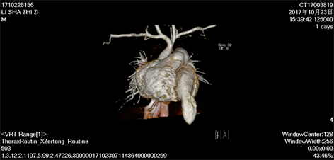

Figure 3. Three dimensional CT imaging of the heart: right deviation of the heart, atrial deficiency, patent ductus arteriosus, and left ventricular diverticulum
图3. 患儿心脏三维CT成像:心脏右旋位,房缺,动脉导管未闭,左心室憩室

Table 1. Left ventricular systolic function test [heart: radial line (mm), area (cm2), velocity (m/s), pressure (mmHg), volume (ml)]
表1. 左心室收缩功能测值(心脏:径线mm,面积cm2;速度m/s,压力mmHg,容量ml)
患儿胸骨缺损、心包部分缺损、膈肌前部缺损、脐上腹壁中线缺如伴心脏膨出、心血管畸形等5种特征,先天性心脏病,为Cantrell五联症。患儿母亲妊娠期有糖尿病、孕早期有感冒,无肝炎、结核及心脏病等疾病,无家族遗传及代谢性疾病,患儿发病原因和发生机制不明,有待进一步研究。
致谢
本病例研究得以报道,首先要感谢患儿家属的知情同意,积极配合,提供给我们非常宝贵的临床资料。其次,感谢新生儿科全体医生的耕耘付出,及时诊断和干预治疗。最后,感谢医院领导对科研的重视与支持。
基金项目
四川省科技厅应用基础计划项目(编号:2016JY0126)。
文章引用
高淑强,肖甜甜,曾雯,巨容. Cantrell五联症新生儿一例并文献复习
Pentalogy of Cantrell Newborn: A Case Report and Literature Review[J]. 亚洲儿科病例研究, 2018, 06(01): 1-5. http://dx.doi.org/10.12677/ACRP.2018.61001
参考文献 (References)
- 1. Cantrell, J.R., Haller, J.A. and Ravitch, M.M. (1958) A Syndrome of Congenital Defects Involving the Abdominal Wall, Sternum, Diaphragm, Pericardium, And Heart. Surgery Gynecology & Obstetrics, 107, 602.
- 2. Korver, A.M.H., Haas, F., Freund, M.W. and Strengers, J.L.M. (2008) Pentalogy of Cantrell. Pediatric Cardiology, 29, 146-149.
- 3. Toyama, W.M. (1972) Combined Congenital Defects of the Anterior Abdominal Wall, Sternum, Diaphragm, Pericardium, and Heart: A Case Report and Review of the Syndrome. Pediatrics, 50, 778.
- 4. Bonilla-Musoles, F., Machado, L.E., Bailão, L.A., Osborne, N.G. and Raga, F. (2001) Abdominal Wall Defects: Two-Versus Three-Dimensional Ultrasonographic Diagnosis. Journal of Ultrasound in Medicine Official Journal of the American Institute of Ultrasound in Medicine, 20, 379-389. https://doi.org/10.7863/jum.2001.20.4.379
- 5. Pirasteh, A., Carcano, C., Kirsch, J. and Mohammed, T.L. (2014) Pentalogy of Cantrell with Ectopia Cordis: CT Findings. Journal of Radiology Case Reports, 8, 29-34. https://doi.org/10.3941/jrcr.v8i12.1972
- 6. 赵迪, 安海燕. Cantrell五联症的诊治进展[J]. 中国全科医学, 2016, 19(14): 1734-1736.
- 7. Kaouthar, H., Jihen, A., Faten, J., Hela, M., Fatma, O., Lilia, C. and Rafik, B. (2013) Cardiac Anomalies in Cantrell’s Pentalogy: From Ventricular Diverticulum to Complete Thoracic Ectopia Cordis. Cardiologie Tunisienne, 9, 94.
- 8. 张学勤, 张伟, 肖明第, 贾宝成, 万亚红, 王吉祥. Cantrell五联症八例的外科诊断与治疗[J]. 中国心血管病研究2011, 9(5): 335-339.
- 9. Türkçapar, A.F., Oruc, A.S., Öksüzoglu, A. and Danışman, N. (2015) Diagnosis of Pentalogy of Cantrell in the First Trimester Using Transvaginal Sonography and Color Doppler. Case Reports in Obstetrics & Gynecology, 2015, 179298;
- 10. Parvari, R., Carmi, R., Weissenbach, J., Pilia, G., Mumm, S. and Weinstein, Y. (1996) Refined Genetic Mapping of X-Linked Thoracoabdominal Syndrome. American Journal of Medical Genetics Part A, 61, 401-402. https://doi.org/10.1002/(SICI)1096-8628(19960202)61:4<401::AID-AJMG18>3.0.CO;2-W