Asian Case Reports in Oncology
Vol.
09
No.
01
(
2020
), Article ID:
34320
,
3
pages
10.12677/acrpo.2020.91001
Primary Gastric Squamous Cell Carcinoma: A Case Report
Yunbo Zhang, Jianguang Zhang, Liping Zheng
Department of Oncology, Zibo Bashan Wanjie Hospital, Zibo Shandong

Received: Feb. 7th, 2020; accepted: Feb. 19th, 2020; published: Feb. 26th, 2020

ABSTRACT
Primary gastric squamous cell carcinoma is a rare disease of the stomach, especially in women. The pathogenesis of gastric squamous cell carcinoma is still unclear. A female patient with gastric squamous cell carcinoma is reported in this paper.
Keywords:Squamous Cell Carcinoma, Primary

原发性胃鳞癌1例
张云波,张建光,郑丽萍
淄博岜山万杰医院肿瘤科,山东 淄博

收稿日期:2020年2月7日;录用日期:2020年2月19日;发布日期:2020年2月26日

摘 要
原发性胃鳞癌(squamous cell carcinoma, SCC)是胃部少见疾病,女性更为罕见。发病机制尚不明确,本文报道一例女性胃鳞癌患者。
关键词 :鳞癌,原发性

Copyright © 2020 by author(s) and Hans Publishers Inc.
This work is licensed under the Creative Commons Attribution International License (CC BY 4.0).
http://creativecommons.org/licenses/by/4.0/


1. 引言
胃癌是全球第四癌症死亡原因,在中国发病率和死亡率居第三位。约50%~70%患者就诊时为进展期 [1]。胃癌最常见的病理类型为腺癌,占90% [2]。鳞癌相对较少,女性患者更为罕见。文献报道也不多。本例患者为我科收治的胃鳞癌患者。该病例经过伦理委员会和患者本人同意。
2. 病例特点
患者女性,67岁,“反复上腹部胀痛不适3月,加重2天。”入院。行14C呼气试验提示HP阳性。胃镜检查:胃底体粘膜粗糙充血,胃底散在斑片状浅糜烂,胃体下部大弯侧见一枚约0.3 × 0.3 cm扁平状息肉,表面光滑,灰白色,给予行高频电切、电凝止血治疗并取活检。胃窦粘膜粗糙,前壁大弯侧可见一处约2.0 × 3.0 cm不规则形凹陷性溃疡,表面粗糙,被覆白苔,周围粘膜隆起,局部取活检。镜下诊断:1) 胃体息肉电切、电凝治疗后;2) 胃窦溃疡(性质待诊);3) 糜烂性胃炎。病理诊断(胃窦大弯侧活检)组织四块,三块低分化癌,根据免疫组化考虑为低分化鳞状细胞癌,一块粘膜组织中度慢性炎伴较多淋巴细胞增生。免疫组化:CK7 (+),CK20 (−),CK5/6 (+),P40 (+),Syn (−),CgA (−),ki-67 > 50% (+) (图1)。行PET-CT排除了其他来源。
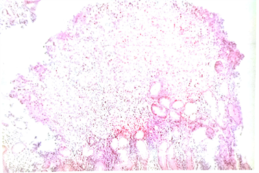
Figure 1. HE, 4X, Greater curvature of antrum
图1. HE、4X、胃窦大弯侧
3. 讨论
原发性胃鳞癌较罕见,发病率在原发性胃癌中大约占0.04%~0.7%,且男性多见,男女之比大约5:1 [3]。Caixia Dong等报道在1988年到2012年间SEER (Surveillance, Epidemiology, and End Results Program)数据库登记的110365例胃癌患者,胃原发鳞状为264例,占原发胃癌的2% [4]。董淑晓等报告了原发性胃鳞癌11例,其中女性仅为3例,且单纯鳞癌为3例,大部分合并腺癌或类癌成分 [5]。临床症状跟腺癌无明显特异性,胃鳞癌诊断主要靠病理学,通常分三个亚型:1) 单纯鳞癌,2) 腺鳞癌(混合型),3) 腺鳞癌(碰撞型) [6]。诊断参照Boswell标准 [7]。Boswell和Hewing定义了4项标准:1) 具有典型癌珠形成的角化细胞团;2) 细胞镶嵌排列,胞界清楚,有单个细胞角化,或细胞核扁而长,胞浆淡伊红染,呈致密的螺旋状排列,表面有早期癌珠形成;3) 有细胞间桥;4) 组化证实癌细胞内有高浓度的硫氢基或二硫基团,表面有角蛋白或前角蛋白存在 [8]。Parks提出发生在贲门和食管胃结合处的不能归为原发性胃鳞癌 [9]。还要除外像肺、气管、宫颈等其他器官鳞癌转移至胃。胃SCC治疗同胃腺癌。一些研究报道病灶较大或明显浸润性生长显示预后较差。然而,另一些研究认为这种类型肿瘤生长缓慢,呈息肉样外生型生长,较腺癌预后好 [4]。Thomas和Sobin应有SEER数据库报告腺癌和鳞癌5年生存率分别为17.1%和13.2%。两种类型结果相似 [10]。
该患者为女性,更为少见。复习近十年文献报道,多为男性患者。多数文献为个案报道。病理由我院诊断,并通过远程会诊均诊断为低分化鳞癌。行PET-CT排除了其他部位来源,发现腹腔淋巴结转移。根据AJCC分期为胃鳞癌IIB期(T3N1M0)。本例患者给予应用阿莫西林 + 克拉霉素 + 奥美拉唑 + 枸橼酸铋钾抗幽门螺杆菌治疗。治疗方案报道无统一标准,主要治疗以手术为主,应用DCF方案术前化疗。治疗反应及预后还需要进一步观察。
基金项目
国家重点研发计划项目(2018YFE0114100)。
文章引用
张云波,张建光,郑丽萍. 原发性胃鳞癌1例
Primary Gastric Squamous Cell Carcinoma: A Case Report[J]. 亚洲肿瘤科病例研究, 2020, 09(01): 1-3. https://doi.org/10.12677/acrpo.2020.91001
参考文献
- 1. Torre, L.A., Bray, F., Siegel, R.L., Ferlay, J., Lortet-Tieulent, J. and Jemal, A. (2015) Global Cancer Statistics, 2012. CA: A Cancer Journal for Clinicians, 65, 87-108. https://doi.org/10.3322/caac.21262
- 2. Thomas, R.M. and Sobin, L.H. (1995) Gastrointestinal Cancer. Cancer, 75, 154-170.
- 3. Dursun, M., Yaldiz, M., Lsikdogan, A., et al. (2003) Primary Squamous Cell Carcinoma of the Stomach: A Case Report and Review of the Literature. European Journal of Gastroenterology & Hepatology, 15, 329-330. https://doi.org/10.1097/00042737-200303000-00018
- 4. Dong, C.X., Jiang, M.J., Tan, Y.N., Kong, Y.Y., Yang, Z.R., Zhong, C.H., et al. (2016) The Clinicopathological Features and Prognostic Factors of Gastric Squamous Cell Carcinoma. Medicine, 95, e4720. https://doi.org/10.1097/MD.0000000000004720
- 5. 董淑晓, 訾力, 亓健. 原发性胃鳞癌的临床病理特点[J]. 中国临床医生, 2011, 39(11): 42-43.
- 6. 陈达丰, 周松. 胃鳞癌1例[J]. 中国普通外科杂志, 2004, 13(12): 920.
- 7. Yoshida, K., Manabe, T., Tsunoda, T., et al. (2002) Early Gastric Cancer of Adenosquamous Carcinoma Type Report of a Case and Review of Literature. Hepatogastroentemlogy, 52, 969-974.
- 8. Boswell, J.T. and Helwig, E.B. (1965) Squamous Cell Carcinoma and Adenoacanthoma of Thestomach. Cancer, 18, 181-192. https://doi.org/10.1002/1097-0142(196502)18:2<181::AID-CNCR2820180209>3.0.CO;2-3
- 9. Parks, R.E. (1967) Squamous Neoplasms of the Stomach. American Journal of Roentgenology, 101, 447-449. https://doi.org/10.2214/ajr.101.2.447
- 10. Thomas, R.M. and Sobin, L.H. (1995) Gastrointestinal Cancer. Cancer, 75, 154-170.