Advances in Clinical Medicine
Vol.
12
No.
04
(
2022
), Article ID:
50939
,
9
pages
10.12677/ACM.2022.124531
TOP2A在肝癌中的表达及其与预后的关系研究
贾清玉*,董银英,闫敏,何信佳,于丽,赵园园#
青岛大学附属医院肿瘤放疗科,山东 青岛
收稿日期:2022年3月28日;录用日期:2022年4月22日;发布日期:2022年4月29日

摘要
目的:探讨DNA拓扑异构酶IIα (topoisomerase II alpha, TOP2A)在肝癌中的表达及其对预后的影响。方法:利用Oncomine数据库分析TOP2A基因在肝癌和正常肝组织中的差异表达。用Western blot和qRT-PCR检测TOP2A在收集的肝癌标本和肝癌细胞系中的表达水平。此外,我们还建立了稳定的肝癌细胞系,在肝癌细胞中下调或过表达TOP2A。运用CCK-8法检测该基因表达如何影响细胞增殖能力。借助GEPIA网站对肝癌数据集进行与TOP2A相关的生存分析。结果:肝癌组织或细胞系中TOP2A的表达水平显著高于正常肝组织及细胞;敲除TOP2A表达可降低原本高表达该基因的肝癌细胞的增殖活力,而上调其表达对原本低表达该基因的细胞的增殖有促进作用,且TOP2A表达量高的患者,其无病生存期(DFS)及总生存期(OS)均较短。结论:TOP2A可能参与肝癌的发生发展,可作为新型肝癌预后标志物,为肝癌精准治疗策略选择提供重要依据。
关键词
TOP2A,肝癌,表达,预后

Study on the Expression of TOP2A in Hepatocellular Carcinoma and Its Relationship with Prognosis
Qingyu Jia*, Yinying Dong, Min Yan, Xinjia He, Li Yu, Yuanyuan Zhao#
Department of Oncology Radiotherapy, The Affiliated Hospital of Qingdao University, Qingdao Shandong
Received: Mar. 28th, 2022; accepted: Apr. 22nd, 2022; published: Apr. 29th, 2022

ABSTRACT
Objective: To investigate the expression of DNA topoisomerase II alpha (TOP2A) in hepatocellular carcinoma (HCC) and its effect on prognosis. Methods: The differential expression of TOP2A in hepatocellular carcinoma and normal liver tissues was analyzed by Oncomine database. Then, we used western blot and qRT-PCR to detect the expression level of TOP2A in collected liver cancer samples and liver cancer cell lines. In addition, we also established a set of stable hepatocellular carcinoma cell lines, which down-regulated or overexpressed TOP2A in them. CCK-8 test was operated to detect how the expression of the gene affected the ability of cell proliferation. The survival analysis of liver cancer data set related to TOP2A was carried out with the assistance of GEPIA. Results: Four studies on the expression of TOP2A in HCC tissues and normal tissues (Roessler Liver, Chen Liver, Wurmbach Liver, Roessler Liver 2) were screened from Oncomine database. The expression level of TOP2A in liver cancer group was higher than that in normal tissue group in 4 cohorts. In a cohort of 40 liver cancer and paracancerous tissue samples we collected after radical resection, the expression of TOP2A mRNA was up-regulated in 33 tumor samples and down-regulated in 7 tumor samples. In addition, we also detected the expression of TOP2A in human normal liver cell line and hepatocellular carcinoma cell line. The results showed that the mRNA and protein levels of TOP2A in high metastatic cell lines MHCC97H, HuH7 and LM3 were significantly higher than those in low metastatic cell line Hep3B and normal liver cell line L02. We chose MHCC97H with relatively high expression of TOP2A and Hep3B with relatively low expression of TOP2A for follow-up experiments. To assess the effect of TOP2A on the viability of MHCC97H and Hep3B cells, the absorbance values (OD) of MHCC97H and Hep3B at 450 nm were recorded by CCK8 assay after transfection by the shTOP2A and pcDNA-TOP2A respectively, and the results showed that TOP2A knockout could reduce the viability of MHCC97H cells, while up-regulation of TOP2A expression promoted the proliferation of Hep3B cells (p < 0.01). The patients with high expression of TOP2A had shorter disease-free survival (DFS) and total survival (OS). Conclusion: TOP2A may be involved in the occurrence and development of hepatocellular carcinoma, and can be regarded as a new type of prognostic marker in liver cancer, which provides an important basis for the selection of accurate treatment strategies for this disease.
Keywords:TOP2A, Hepatocellular Carcinoma, Expression, Prognosis

Copyright © 2022 by author(s) and Hans Publishers Inc.
This work is licensed under the Creative Commons Attribution International License (CC BY 4.0).
http://creativecommons.org/licenses/by/4.0/


1. 引言
据WHO统计,在世界范围内,肝癌是癌症相关死亡的第四大原因,在发病率方面排名第六 [1]。肝癌在我国占癌症相关死亡原因的12.9%,在消化道恶性肿瘤中排第二位,在男性最常见的癌症相关死亡原因中位列仅次于肺癌 [2]。根治性手术诊断为肝癌的患者提供了长期生存的唯一希望。然而,当有临床表现时,大多数患者已进入晚期,失去手术机会。临床上预测肝癌预后的指标有限,如肿瘤大小、肝硬化程度、肿瘤分化程度和肿瘤微血管侵犯程度 [3],且暂无研究表明具有明确诊断意义的AFP可以预测肝癌分期及预后。由于肝癌根治性切除术后肿瘤复发率和转移率较高,且肝癌预后缺乏分子层面指标,因此了解与肝癌转移相关的分子机制和寻找新的治疗靶点对肝癌患者的预后或具有重要意义。
DNA拓扑异构酶是一种解开双链DNA中出现的拓扑问题的酶 [4]。根据氨基酸序列结构和/或关系的不同,DNA拓扑异构酶又分为:IA (DNA拓扑异构酶IIIα和IIIβ)、IB (DNA拓扑异构酶I和线粒体DNA拓扑异构酶I99)和IIA (DNA拓扑异构酶IIα和IIβ) [5]。其中DNA拓扑异构酶IIα (TOP2A)参与染色体凝聚、染色单体分离以及减轻DNA转录和复制过程中产生的扭应力等过程。它通过催化两条双链DNA的瞬间断裂和重新连接,从而允许这两条链相互穿透,从而改变DNA的拓扑结构。此外,TOP2A在快速分裂的细胞中大量积累,其表达与细胞增殖和细胞周期有关,其产生和降解受细胞周期调控 [6] [7] [8]。
2. 材料和方法
2.1. 数据筛选
利用Oncomine数据库(http://www.oncomine.org)分析肝癌患者血清中TOP2A的表达水平,通过比较肝癌组织数据集和正常组织数据集探索TOP2A更多信息。
2.2. 肿瘤组织标本选择
本研究收集了40例接受根治性肝癌切除术患者肿瘤标本和配对的癌旁正常肝标本。本研究已得到复旦大学中山医院(中国上海)研究伦理委员会批准(鉴定代码:B2013-150)。
2.3. 细胞培养
5个肝癌细胞系(Hep3B、LM3、Huh7、MHCC97H)和1个正常人肝细胞系(L02)均置于DMEM培养基中且培养在37℃、5% CO2的培养箱环境内。
2.4. RNA提取和qRT-PCR
按照Trizol试剂说明书分别提取组织及细胞样本总RNA。并按照Takara试剂盒(大连宝生物)将提取出的RNA逆转录为cDNA,以95℃预变性30 s;95℃ 5 s,60℃ 30 s,共40个循环;95℃ 5 s,60℃ 1 min的程序扩增cDNA,并用Light Cycler 480 II (罗氏)对其进行检测。以GAPDH为内源性参照物,用2−ΔΔCt法测定相对基因表达。所用引物序列如表1所示。

Table 1. Primer sequence used for qRT-PCR
表1. qRT-PCR 反应引物序列
2.5. Western Blot
用含有RIPA (碧云天)、PMSF (碧云天)和10% PhosSTOP的混合裂解缓冲液从肝癌细胞中提取总蛋白,并用10%的SDS-PAGE分离,然后转移到PVDF膜上。室温下用5%脱脂牛奶在TBST缓冲液中封闭膜1 h,然后在4℃条件下与稀释的一抗(1:1000, Abcam)反应过夜。进一步将膜与HRP偶联的二抗(1:1000,北京鼎国昌盛生物)在室温下孵育1 h。最后,使用电化学发光试剂盒(Thermo)使目标条带显影。
2.6. 慢病毒构建与细胞转染
pGCSIL-shRNA-TOP2A慢病毒载体和pGC-FU-TOP2A cDNA慢病毒载体购自上海GeneChem有限公司。敲除TOP2A最有效的shRNA慢病毒载体序列是5’-ATCCTGCAG-GAATGGCATT-3’,同时设置无特异性转染的阴性对照,其序列为5’-TTCTCCGAACGTGTCACGT-3’。以pGC-FU慢病毒载体为参照,构建了可上调TOP2A表达的pcDNA3.1载体。通过qRT-PCR和Western blot分析对稳定转染的克隆进行验证。
2.7. CCK-8增殖试验
将100 µL细胞悬液(约2 × 10⁴/ml)加入至培养板孔。细胞贴壁后,每孔加入10 µL CCK-8溶液,在培养箱中孵育1 h。用酶标仪测定细胞在450 nm处的吸光度值(OD)。空白对照为加入等量不含细胞的细胞培养液和CCK-8溶液。
2.8. 生存分析
在基因表达谱交互分析(GEPIA)网站(http://gepia.cancer-pku.cn/index.html) [9] 上进行肝细胞癌TOP2A数据集的生存分析。检索条件:1) Gene:TOP2A;2) Methods:OS OR DFS;3) Group Cutoff:Median;4) Hazards Ratio (HR):Yes;5) 95% Confidence Interval:Yes;6) Axis Units:Months;7) Datasets Selection (Cancer name):LIHC。
2.9. 统计分析
采用GraphPad Prism 5软件进行统计分析。两组间的差异用t检验进行分析,组间差异比较采用单因素方差分析(ANOVA)检验。每个实验至少重复三次,测量数据用均值 ± 标准差表示。P < 0.05具有统计学意义。
3. 结果
3.1. TOP2A在人肝癌细胞和组织中表达上调
在Oncomine数据库中筛选了4项有关TOP2A在HCC组织和正常组织中表达的研究(Roessler Liver, Chen Liver, Wurmbach Liver, Roessler Liver 2),共有712个样本。如图1(A)所示,4个研究队列中肝癌组织组TOP2A的表达水平均高于正常组织组(p < 0.05)。另外,在一个由我们收集的40个连续行根治术后取得的肝癌组织及癌旁组织样本组成的队列中,经qRT-PCR检测,33个肿瘤样本(82.5%)中TOP2A mRNA表达水平呈上调状态,7个肿瘤样本(17.5%)中TOP2A mRNA表达下调(图1(B))。这一结果在从该队列中随机选择的8对肝癌(T)及其癌旁正常组织(N)样本中用Western blot方法进行了验证,如图1(C)所示。此外,我们还检测了TOP2A在人正常肝细胞系及肝癌细胞系中的表达。结果显示,TOP2A在高转移细胞系MHCC97H、HuH7和HCCLM3的mRNA和蛋白水平均显著高于低转移细胞系Hep3B和正常肝细胞系L02 (见图1(D)),**p < 0.01。在此我们选择相对高表达TOP2A的MHCC97H和相对低表达TOP2A的Hep3B进行后续实验。
3.2. TOP2A促进肝癌细胞体外增殖
为了探究敲低或上调TOP2A表达后对肝癌细胞增殖情况的影响,我们将shTOP2A-1、shTOP2A-2、shTOP2A-Ctrl分别转染至原本高表达TOP2A的MHCC97H细胞中,并用Western Blot检测转染效果,图示shTOP2A-2转染效率较高;同时将含pcDNA-TOP2A及pcDNA-Ctrl的质粒分别转染原本低表达TOP2A的Hep3B细胞,Western Blot结果示上调基因表达后TOP2A蛋白表达明显增加(图2(A))。另外用qRT-PCR验证了shRNA在MHCC97H细胞中敲除TOP2A的效率和pcDNA在Hep3B细胞中上调TOP2A的结果(见图2(B)),***p < 0.001。为评估TOP2A对MHCC97H和Hep3B细胞活性的影响,在shTOP2A或pcDNA-TOP2A转染0 h、12 h、24 h、48 h、72 h后采用CCK8分析法分别记录MHCC97H和Hep3B在450 nm处的吸光度值(OD),如图2(C)所示,TOP2A基因敲除可降低MHCC97H细胞的活力,而上调TOP2A表达对Hep3B细胞的增殖有促进作用,**p < 0.01。

Figure 1. (A) Expression levels of TOP2A in normal liver tissues and liver cancer tissues in four studied arrays from the Oncomine database. (B) qRT-PCR was performed on TOP2A mRNA expression in tumor tissues from 40 patients who underwent curative resection of liver cancer. Expression of (C) TOP2A protein in normal tissues (N) and hepatocellular carcinoma tissues (T). Expression of (D) TOP2A mRNA and protein in human normal liver cell line L02 and hepatoma cell lines Hep3B, LM3, Huh7, MHCC97H. *p < 0.05, **p < 0.01
图1. (A) 来自Oncomine数据库的4个研究阵列中正常肝脏组织及肝癌组织中TOP2A的表达水平。(B) 对40名接受肝癌根治性切除术患者的肿瘤组织中TOP2A mRNA表达进行qRT-PCR检测。(C) TOP2A蛋白在癌旁正常组织(N)和肝癌组织(T)中的表达。(D) TOP2A mRNA和蛋白人正常肝细胞系L02和肝癌细胞系Hep3B、LM3、Huh7、MHCC97H中的表达。*p < 0.05,**p < 0.01
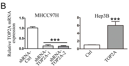


Figure 2. (A) TOP2A protein expression levels in MHCC97H or Hep3B cells after knockdown or up-regulation of TOP2A gene expression. (B) qRT-PCR was performed to detect the knockdown efficiency of TOP2A in MHCC97H cells by shRNA and the stable up-regulation of TOP2A expression in Hep3B cells by pcDNA, respectively. ***p < 0.001. (C) The effect of knockdown or up-regulation of TOP2A gene expression on the proliferation of MHCC97H or Hep3B cells was analyzed by CCK8 assay. **p < 0.01
图2. (A) 敲除或上调TOP2A基因表达后MHCC97H或Hep3B细胞TOP2A蛋白表达水平。(B) qRT-PCR分别检测shRNA在MHCC97H细胞中TOP2A的敲除效率和pcDNA使Hep3B细胞中TOP2A表达的稳定上调。***p < 0.001。(C) CCK8法分析敲低或上调TOP2A基因表达对MHCC97H或Hep3B细胞增殖的影响。**p < 0.01
3.3. TOP2A表达水平与肝癌患者预后相关
GEPIA (Gene Expression Profile Interactive Analysis)是一个基于Web的工具,可以根据TCGA和GTEx数据提供关键的交互式和可定制功能,包括差异表达分析、轮廓绘制、相关性分析、患者生存分析、相似基因检测和降维分析 [9]。本研究借助GEPIA内数据库进行生存分析(图3(A)、图3(B)),结果显示高表达TOP2A的患者DFS及OS均劣于低表达患者。

Figure 3. The level of (A) TOP2A expression correlated with disease-free survival (DFS). (B) TOP2A expression levels correlate with overall survival (OS)
图3. (A) TOP2A表达水平与无病生存期(DFS)相关。(B) TOP2A表达水平与总生存期(OS)相关
4. 讨论
肝癌是全世界最常见的恶性肿瘤之一。经过多年的发展,肝癌的治疗方法已从早期的手术切除、肝移植、TACE、化疗和放疗逐渐演变为分子靶向治疗结合免疫治疗 [10]。AFP先前被指南推荐作为监测HCC的血清标志物。Agopian等人 [11] 的一项研究报告说,在665名HCC患者中,31.3%的AFP水平在正常范围内。虽然直到现在AFP仍在临床上使用,但它不作为监测指标用于AFP水平正常的患者 [12]。欧洲肝脏研究协会(EASL)建议,除了甲胎蛋白,血管内皮生长因子和血管生成素2也可以作为预后标记物 [13]。此外,寻找新的有效的肝癌预后生物标志物的努力正在进行中。
TOP2A在不同类型肿瘤中被检测到高表达,如肺癌 [14] [15]、结肠癌 [16] [17]、膀胱尿路上皮癌 [18]、前列腺癌 [19] [20]、乳腺癌 [21] [22]、卵巢癌 [23] 等,均提示与不良预后相关。Panvichian等人 [24] 证实TOP2A在HCC中的高表达与Ki-67的高表达有关,Ki-67的表达与HCC的肿瘤生长速度和不良预后有关 [25],提示TOP2A可以作为治疗肝癌的潜在靶点。我们的研究首先验证了肝癌组织和高转移肝癌细胞系中TOP2A在mRNA和蛋白水平表达均高于正常组织和低转移细胞系。TOP2A主要位于处在分裂增殖状态细胞的细胞核内,且在快速增殖的细胞中,其表达水平是静止细胞的数倍。在细胞周期中,TOP2A在G0/G1期含量较低,S期增多,G2/M期达到高峰,它在此期间发挥调控核酸空间结构动态变化、调控核酸生理功能的关键作用 [26] [27]。我们通过慢病毒载体稳定转染构建了过表达和敲除TOP2A的两组细胞,通过CCK8试验发现上调TOP2A基因表达后低转移细胞系增殖能力明显增加,而敲除该基因后高转移细胞系增殖能力相应下降(p < 0.01)。这与前人的部分研究 [28] 结果相符,提示TOP2A在肝癌组织中的高表达可能与肿瘤的发展有关。
根据GEPIA数据库显示,高表达TOP2A的患者OS和DFS均高于低表达TOP2A的患者。CAI [26] 等人的研究中发现,与白人种族相比,亚洲人群中TOP2A的高表达与不良预后之间的关联更为显著。这可能与亚洲肝癌的病因多与病毒性肝炎有关,而肝炎病毒又对DNA复制过程产生影响。本研究在生存分析方面存在一定局限性。虽然GEPIA是获得研究线索的有用资源,但这一结果后续需要在大量临床样本中进行验证。
5. 结论
综上所述,我们的研究结果验证了相较于正常细胞和组织,TOP2A在肝癌细胞和组织中呈现高表达水平,表明TOP2A可能与部分肝癌的发生有关。而且在上调TOP2A基因表达时会提高原本低扩增的细胞系的增殖能力,相反,下调其表达会减少高扩增细胞系的增殖,这表明TOP2A的表达水平可能与肝癌细胞倍增有关。检索GEPIA数据库显示,当以OS和DFS为观察终点时,TOP2A高表达的患者预后差于低表达患者。因此TOP2A将来有潜力成为预测肝癌预后的重要生物标志物。
文章引用
贾清玉,董银英,闫 敏,何信佳,于 丽,赵园园. TOP2A在肝癌中的表达及其与预后的关系研究
Study on the Expression of TOP2A in Hepatocellular Carcinoma and Its Relationship with Prognosis[J]. 临床医学进展, 2022, 12(04): 3668-3676. https://doi.org/10.12677/ACM.2022.124531
参考文献
- 1. Villanueva, A. (2019) Hepatocellular Carcinoma. The New England Journal of Medicine, 380, 1450-1462. https://doi.org/10.1056/NEJMra1713263
- 2. Feng, R., Zong, Y., Cao, S. and Xu, R.-H. (2019) Current cancer situation in China: good or bad news from the 2018 Global Cancer Statistics? Cancer Communications, 39, Article No. 22. https://doi.org/10.1186/s40880-019-0368-6
- 3. Sharma, S., Kowgier, M., Hansen, B., Brouwer, W.P., Maan, R., Wong, D., et al. (2018) Toronto HCC Risk Index: A Validated Scoring System to Predict 10-Year Risk of HCC in Patients with Cirrhosis. Journal of Hepatology, 68, 92-99. https://doi.org/10.1016/j.jhep.2017.07.033
- 4. Nitiss, J.L. (2009) DNA Topoisomerase II and Its Growing Repertoire of Biological Functions. Nature Reviews Cancer, 9, 327-37. https://doi.org/10.1038/nrc2608
- 5. Wang, J.C. (2002) Cellular Roles of DNA Topoisomerases: A Molecular Perspective. Nature Reviews Molecular Cell Biology, 3, 430-440. https://doi.org/10.1038/nrm831
- 6. Erguden, H., Koksal, D., Demirag, F., Bayiz, H., Mutluay, N., Berktas, B., et al. (2012) The Association of Topoisomerase 2α Expression with Prognosis in Surgically Resected Non-Small Cell Lung Cancer (NSCLC) Patients. Journal of Thoracic Disease, 4, 352-357.
- 7. Demoulin, B., Hermant, M., Castrogiovanni, C., Staudt, C. and Dumont, P. (2015) Resveratrol Induces DNA Damage in Colon Cancer Cells by Poisoning Topoisomerase II and Activates the ATM Kinase to Trigger p53-Dependent Apoptosis. Toxicology in Vitro, 29, 1156-1165. https://doi.org/10.1016/j.tiv.2015.04.015
- 8. Sudan, S. and Rupasinghe, H.P. (2014) Flavonoid-Enriched Apple Fraction AF4 Induces Cell Cycle Arrest, DNA Topoisomerase II Inhibition, and Apoptosis in Human Liver Cancer HepG2 Cells. Nutrition and Cancer, 66, 1237-1246. https://doi.org/10.1080/01635581.2014.951733
- 9. Tang, Z., Li, C., Kang, B., Gao, G., Li, C. and Zhang, Z. (2017) GEPIA: A Web Server for Cancer and Normal Gene Expression Profiling and Interactive Analyses. Nucleic Acids Research, 45, W98-W102. https://doi.org/10.1093/nar/gkx247
- 10. Yau, T., Hsu, C., Kim, T., Choo, S.P., Kang, Y.K., Hou, M.M., et al. (2019) Nivolumab in Advanced Hepatocellular Carcinoma: Sorafenib-Experienced Asian Cohort Analysis. Journal of Hepatology, 71, 543-552. https://doi.org/10.1016/j.jhep.2019.05.014
- 11. Agopian, V., Harlander-Locke, M., Markovic, D., Zarrinpar, A., Kaldas, F.M., Cheng, E.Y., et al. (2017) Evaluation of Patients With Hepatocellular Carcinomas That Do Not Produce α-Fetoprotein. JAMA Surgery, 152, 55-64. https://doi.org/10.1001/jamasurg.2016.3310
- 12. Luo, P., Wu, S., Yu, Y., Ming, X., Li, S., Zuo, X., et al. (2020) Current Status and Perspective Biomarkers in AFP Negative HCC: Towards Screening for and Diagnosing Hepatocellular Carcinoma at an Earlier Stage. Pathology Oncology Research, 26, 599-603. https://doi.org/10.1007/s12253-019-00585-5
- 13. Tsochatzis, E., Meyer, T., O’Beirne, J., et al. (2013) Transarterial Chemoembolisation Is Not Superior to Embolisation Alone: The Recent European Association for the Study of the Liver (EASL)—European Organization for Research and Treatment of Cancer (EORTC) Guidelines. European Journal of Cancer, 49, 1509-1510. https://doi.org/10.1016/j.ejca.2012.11.012
- 14. Kou, F., Sun, H., Wu, L., Li, B., Zhang, B., Wang, X., et al. (2020) TOP2A Promotes Lung Adenocarcinoma Cells’ Malignant Progression and Predicts Poor Prognosis in Lung Adenocarcinoma. Journal of Cancer, 11, 2496-508. https://doi.org/10.7150/jca.41415
- 15. Dingemans, A.M., Witlox, M.A., Stallaert, R.A., van der Valk, P., Postmus, P.E. and Giaccone, G. (1999) Expression of DNA Topoisomerase IIalpha and Topoisomerase IIbeta Genes Predicts Survival and Response to Chemotherapy in Patients with Small Cell Lung Cancer. Clinical Cancer Research, 5, 2048-2058.
- 16. Lazaris, A.C., Kavantzas, N.G., Zorzos, H.S., Tsavaris, N.V. and Davaris, P.S. (2002) Markers of Drug Resistance in Relapsing Colon Cancer. Journal of Cancer Research and Clinical Oncology, 128, 114-118. https://doi.org/10.1007/s00432-001-0310-5
- 17. Zhang, R., Xu, J., Zhao, J. and Bai, J.H. (2018) Proliferation and Invasion of Colon Cancer Cells Are Suppressed by Knockdown of TOP2A. Journal of Cellular Biochemistry, 119, 7256-7263. https://doi.org/10.1002/jcb.26916
- 18. Zeng, S., Liu, A., Dai, L., Yu, X., Zhang, Z., Xiong, Q., et al. (2019) Prognostic Value of TOP2A in Bladder Urothelial Carcinoma and Potential Molecular Mechanisms. BMC Cancer, 19, Article No. 604. https://doi.org/10.1186/s12885-019-5814-y
- 19. Nelson, W.G., Haffner, M.C. and Yegnasubramanian, S. (2018) The Structure of the Nucleus in Normal and Neoplastic Prostate Cells: Untangling the Role of Type 2 DNA Topoisomerases. American Journal of Clinical and Experimental Urology, 6, 107-113.
- 20. De Resende, M.F., Vieira, S., Chinen, L.T., Chiappelli, F., da Fonseca, F.P., Guimarães, G.C., et al. (2013) Prognostication of Prostate Cancer Based on TOP2A Protein and Gene Assessment: TOP2A in Prostate Cancer. Journal of Translational Medicine, 11, Article No. 36. https://doi.org/10.1186/1479-5876-11-36
- 21. Fritz, P., Cabrera, C.M., Dippon, J., Gerteis, A., Simon, W., Aulitzky, W.E., et al. (2005) c-erbB2 and Topoisomerase IIalpha Protein Expression Independently Predict Poor Survival in Primary Human Breast Cancer: A Retrospective Study. Breast Cancer Research, 7, Article No. R374. https://doi.org/10.1186/bcr1012
- 22. Badawy, O.M. and Loay, I. (2019) FISH Analysis of TOP2A and HER-2 Aberrations in Female Breast Carcinoma on Archived Material: Egyptian NCI Experience. Applied Immunohistochemistry & Molecular Morphology, 27, 216-222. https://doi.org/10.1097/PAI.0000000000000574
- 23. Costa, M.J., Hansen, C.L., Holden, J.A. and Guinee, D. (2000) Topoisomerase II Alpha: Prognostic Predictor and Cell Cycle Marker in Surface Epithelial Neoplasms of the Ovary and Peritoneum. International Journal of Gynecological Pathology, 19, 248-257. https://doi.org/10.1097/00004347-200007000-00009
- 24. Panvichian, R., Tantiwetrueangdet, A., Angkathunyakul, N. and Leelaudomlipi, S. (2015) TOP2A Amplification and Overexpression in Hepatocellular Carcinoma Tissues. BioMed Research International, 2015, Article ID: 381602. https://doi.org/10.1155/2015/381602
- 25. Ng, I., Na, J., Lai, E., Fan, S.T. and Ng, M. (1995) Ki-67 Antigen Expression in Hepatocellular Carcinoma Using Monoclonal Antibody MIB1. A Comparison with Proliferating Cell Nuclear Antigen. American Journal of Clinical Pathology, 104, 313-318. https://doi.org/10.1093/ajcp/104.3.313
- 26. Cai, H., Zhu, X., Qian, F., Shao, B., Zhou, Y., Zhang, Y., et al. (2020) High Expression of TOP2A Gene Predicted Poor Prognosis of Hepatocellular Carcinoma after Radical Hepatectomy. Translational Cancer Research, 9, 983-992. https://doi.org/10.21037/tcr.2019.12.46
- 27. Anand, J., Sun, Y., Zhao, Y., Nitiss K.C. and Nitiss J.L. (2018) Detection of Topoisomerase Covalent Complexes in Eukaryotic Cells. In: Drolet, M., Eds., DNA Topoisomerases, Vol. 1703, Humana Press, New York, 283-299. https://doi.org/10.1007/978-1-4939-7459-7_20
- 28. Wong, N., Yen, W., Wong, W.-L., Wong, N.L.-Y., Chan, K.Y.-Y., Mo, F.K.-F., et al. (2009) TOP2A Overexpression in Hepatocellular Carcinoma Correlates with Early Age Onset, Shorter Patients Survival and Chemoresistance. International Journal of Cancer, 124, 644-652. https://doi.org/10.1002/ijc.23968
NOTES
*第一作者。
#通讯作者Email: 18661807696@163.com