Advances in Clinical Medicine
Vol.
13
No.
10
(
2023
), Article ID:
73613
,
5
pages
10.12677/ACM.2023.13102221
胆囊炎性肌纤维母细胞瘤1例
张晓菡1,王晓艳2,熊浩1*
1长江大学附属第一医院放射科,湖北 荆州
2长江大学附属第一医院病理科,湖北 荆州
收稿日期:2023年9月11日;录用日期:2023年10月5日;发布日期:2023年10月12日

摘要
目的:通过病例回顾,提高对胆囊炎性肌纤维母细胞瘤的认识、诊断和治疗。方法:总结1例经病理证实的胆囊炎性肌纤维母细胞瘤患者的临床资料,并复习相关文献资料。结果:患者临床表现为右上腹、剑突下隐痛。CT示胆囊壁增厚,局部可见片状软组织密度影,增强扫描呈中度强化,局部与肝实质界限不清。MR示胆囊颈部可见类圆形短T2信号影,胆囊底部可见不规则团块状异常信号影,以稍长T1稍长T2信号为主,边界清晰,DWI呈稍高信号影,增强呈明显延迟强化,病灶与邻近肝实质界限不清。胆囊病灶术后病理确诊为胆囊炎性肌纤维母细胞瘤伴坏死。结论:胆囊炎性肌纤维母细胞瘤属于良性肿瘤,其在胆囊中的发生率极低,临床表现不典型,影像学缺乏特异性,确诊仍依赖于病理。手术为其主要的治疗方法,但易复发,因此应加强对其随访。
关键词
炎性肌纤维母细胞瘤,胆囊,CT,MRI

A Case Report of Inflammatory Myofibroblastic Tumor of the Gallbladder
Xiaohan Zhang1, Xiaoyan Wang2, Hao Xiong1*
1Department of Radiology, The First Affiliated Hospital of Yangtze University, Jingzhou Hubei
2Department of Pathology, The First Affiliated Hospital of Yangtze University, Jingzhou Hubei
Received: Sep. 11th, 2023; accepted: Oct. 5th, 2023; published: Oct. 12th, 2023

ABSTRACT
Objective: To improve the understanding, diagnosis and treatment of inflammatory myofibroblastic tumor (IMT) in gallbladder through the review of the case. Methods: The clinical data of a case of pathologically proved IMT in gallbladder was reported and relevant literatures were reviewed. Results: The patient felt dull pain in the right upper abdomen and below xiphoid. CT showed thickening of the gallbladder wall, local patchy soft tissue density, moderate enhancement with contrast, and unclear local boundary with liver parenchyma. MR showed circular and short T2 signal in the neck of the gallbladder, irregular regiment massive in the bottom of the gallbladder, mainly with slightly long T1 and long T2 signal, with clear boundary; DWI showed slightly high signal, and the enhancement showed obvious delayed enhancement, and the boundary between the lesion and the adjacent liver parenchyma was unclear. The pathological diagnosis of the gallbladder lesion was inflammatory myofibroblastic tumor with necrosis. Conclusion: IMT is a benign tumor and is rare in the gallbladder. The clinical manifestations are not typical and the imaging manifestations are not specific. lts diagnosis depends on pathology. Surgery is the main treatment, but IMT has the tendency of recurrence, so its postoperative follow-up is of great significance.
Keywords:Inflammatory Myofibroblastic Tumor, Gallbladder, CT, MRI

Copyright © 2023 by author(s) and Hans Publishers Inc.
This work is licensed under the Creative Commons Attribution International License (CC BY 4.0).
http://creativecommons.org/licenses/by/4.0/


1. 引言
炎性肌纤维母细胞瘤(inflammatory myofibroblastic tumor, IMT)是近年来逐渐认识的一种少见的间叶肿瘤性疾病,主要由分化的肌纤维母细胞性梭形细胞组成,其发病机制尚不明确,发病率低、复发率高、转移率高,临床症状和影像学表现缺乏特异性,因此手术前诊断困难,易误诊、漏诊 [1] 。笔者回顾性分析1例经病理证实的IMT患者的临床、影像、病理资料,并查阅相关文献,以提高对本病的认识和影像诊断水平。
病例简介:患者女,49岁,4天前无诱因出现右上腹、剑突下隐痛,无反酸,疼痛持续约30分钟左右自行好转,无腹泻、恶心、呕吐、发热,未特殊处理,症状自行好转,但症状反复,常于夜间复发,1天前腹痛再发。患者有2型糖尿病史。体格检查未见明显异常。实验室检查:糖类抗原CA-199:942.88 U/mL,癌胚抗原CEA:6.05 ng/mL。患者自本次发病以来,精神可,胃纳略差,睡眠可,大便如常,小便如常,体重未见明显下降。
辅助检查:1) CT:胆囊壁增厚,局部可见片状软组织密度影,增强扫描呈中度强化,局部与肝实质界限不清(图1)。2) MRI:胆囊不大,胆囊颈部可见类圆形短T2信号影,大小约22 mm × 14 mm;胆囊底部可见不规则团块状异常信号影,以稍长T1稍长T2信号为主,边界清晰,大小约36 mm × 39 mm × 36 mm (上下径 × 前后径 × 左右径),DWI呈稍高信号影,增强呈明显延迟强化,病灶与邻近肝实质界限不清(图2)。
手术:患者行“腹腔镜下胆囊根治性切除术 + 肝部分切除 + 腹腔淋巴清扫胆管修补 + 胆道镜检 + 肠粘连松解”术,术中见胆囊灰白质硬占位累及周围肝实质,侵及胃十二指肠,盆腔腹膜、肠系膜可及扪及多发灰白质硬结节,胰头后方可扪及多发肿大淋巴结。探查腹腔内大网膜与胆囊粘连,胆囊大小约7.5 × 3.5 cm,胆囊底部僵硬明显,与肝脏界限不清,胆囊颈部膨大,其内含2.5 cm大小结石,向腹侧压迫肝总管,肝总管与胆囊颈部粘连。
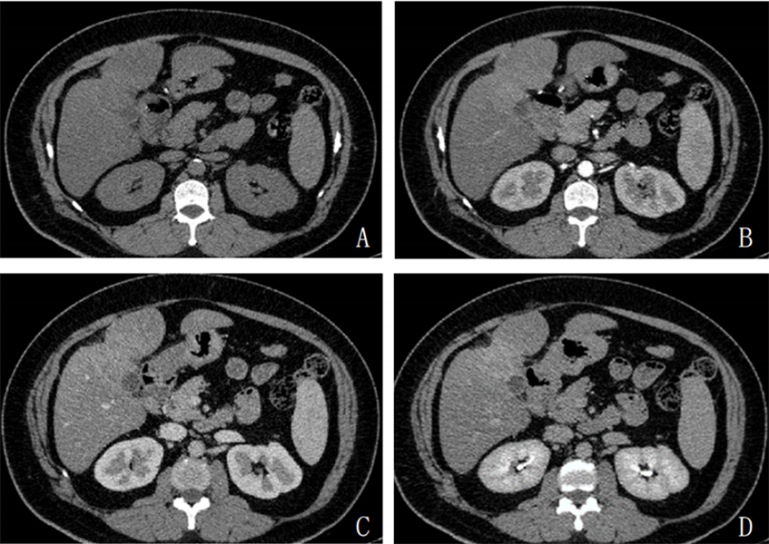
Figure 1. Epigastric CT showed thickening of the gallbladder wall, local patchy soft tissue density, moderate enhancement with contrast, and unclear local boundary with liver parenchyma
图1. 上腹部CT示胆囊壁增厚,局部可见片状软组织密度影,增强扫描呈中度强化,局部与肝实质界限不清
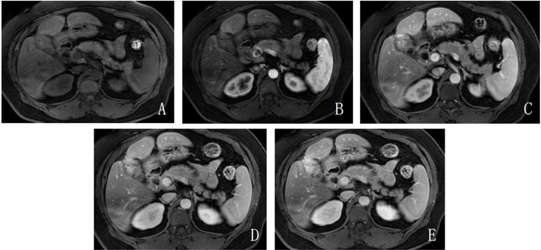
Figure 2. The T1WI axial enhanced scan showed obvious delayed enhancement, and the boundary between the lesion and the adjacent liver parenchyma was unclear
图2. T1WI轴位增强扫描示明显延迟强化,病灶与邻近肝实质界限不清
术后病理:1) 病理标本肉眼所见:胆囊底部质硬,剖开胆囊腔内见黄色2.5 cm结石一枚,底部僵硬增厚明显,与肝脏界限不清。2) 镜下见瘤组织与肝脏粘连,周围较多炎性细胞及多核巨细胞浸润;免疫组化检测:PCK(−),Vimentin(+),hepatocyte(−),Glypican-3(−),CD34血管(+),SMA(+),Desmin少许(+),Calponin(+),S−100(−),CD68(+),ALK(−),Ki67 Li约5%。病理诊断:胆囊炎性肌纤维母细胞瘤伴坏死。
2. 讨论
炎性肌纤维母细胞瘤是一种少见的间叶组织来源的肿瘤,虽具有恶性潜能和复发潜力,但大多为良性 [2] [3] ,极少发生转移。它可以发生在肺、脾、肝、淋巴结、胰腺和肝外胆管等部位,好发于儿童和青年的肺部 [4] 。胆囊原发性炎性肌纤维母细胞瘤相当罕见,目前对它的了解是基于个案报道 [5] 。IMT确诊依靠的是病理,在组织学上,IMT主要由分化的肌纤维母细胞性梭形细胞构成,并伴有浆细胞、淋巴细胞等炎症浸润,在免疫组化中,大部分表达SMA、Vimentin或Desmin [6] [7] [8] 。
炎性肌纤维母细胞瘤曾经被称为炎性假瘤、浆细胞瘤等。IMT首次在肺部观察到,并由Bunn于1939年描述 [9] 。因为其在临床、放射学和组织病理学上模仿恶性肿瘤,所以Umiker等于1954年将其命名为“炎性假瘤” [10] 。但当通过免疫组织化学标记和电子显微镜研究确定其细胞来源为肌纤维母细胞时,便改称为“炎性肌纤维母细胞瘤”。2002年,世界卫生组织国际软组织肿瘤组织学分类专家正式将其命名为炎性肌纤维母细胞瘤(IMT),是指“由分化的肌纤维母细胞性梭形细胞组成的,常伴大量浆细胞和(或)淋巴细胞的一种肿瘤” [11] 。
IMT的基本组成部分为淋巴细胞、浆细胞、组织细胞、成纤维细胞和肌成纤维细胞,这些组成部分比例各不相同。四种常见的基本组织学模式分别为:a) 淋巴浆细胞浸润为主;b) 淋巴组织细胞浸润为主;c) 年轻且活跃的肌成纤维细胞过程;d) 以胶原化为主的过程,伴有淋巴细胞浸润。
IMT的病因至今仍不清楚,但认为主要与以下因素有关:1) 感染:乙型和丙型肝炎病毒等感染性微生物在目标器官释放炎性介质,引起急性渗出性病变,并逐渐纤维化,形成炎性假瘤 [12] 。在这个病例中,胆囊IMT患者在手术前有感染的表现,但还没有明确的证据证明这是引起它发生的原因。2) 手术或创伤所致的自身免疫异常:以往的研究表明,肝脏IMT患者的血清免疫球蛋白IgG4明显升高,并且可以在病灶内找到IgG4阳性的浆细胞 [13] 。但由于缺乏对该病的了解,患者在术前未能完成IgG4水平的测定。3) 基因异常:最近的研究表明,部分IMT与异常基因ALK (也被称为中间变性淋巴瘤激酶 [14] )表达有关。4) 放化疗、过敏等 [15] 。
胆囊的IMT一般没有特异性临床症状,与肿物的部位有一定的关系。本例中胆囊IMT有胆囊炎表现,症状为右上腹、剑突下隐痛。实验室检查结果也缺乏特异性,IMT没有特异性肿瘤标志物 [16] 。糖尿病患者由于容易感染,临床结果非常差,需要特别关注,本例的病人即为2型糖尿病患者 [17] 。
CT和MR检查能显示病灶的形态、内部结构及与周围组织的关系,有助于临床的诊治,但是IMT的影像特征缺乏特异性,与其他病变有相似之处,特别是恶性肿瘤,因而无法作出准确诊断 [18] [19] [20] 。IMT的细胞类型以肌成纤维细胞为主,其最具特异性的特征是具有模仿恶性的能力,肌成纤维细胞增殖越密集,与恶性肿瘤相鉴别就会越困难。IMT定位于胆囊,应与原发性胆管癌、胆囊癌等进行鉴别 [21] 。大部分的病例是根据组织病理学和免疫组织化学研究作出最终诊断 [22] [23] 。此外,组织学检查排除了另一种良性病变:黄色肉芽肿性胆囊炎,这是一种慢性胆囊炎性疾病,可见由广泛的纤维化和泡沫细胞组成的多个黄褐色壁内结节。与IMT类似,这是一种罕见的起源不明疾病,二者的鉴别完全依赖于病理检查。
IMT首选治疗方法为手术全切除 [24] ,术后通常不需要再进行放化疗 [25] 。除了手术治疗,还有其他治疗方式,如激素、抗生素和ALK小分子ATP竞争性抑制剂等,这些治疗方式也有一定的报道。然而,对于无法确诊并且不能排除恶性肿瘤的患者,应积极进行外科治疗。
众所周知,这些肿瘤具有很强的增殖能力,可渗透到周围组织,与周围重要组织黏连未能完全切除,易导致肿瘤复发,因此术后需严密随访 [26] [27] 。
文章引用
张晓菡,王晓艳,熊 浩. 胆囊炎性肌纤维母细胞瘤1例
A Case Report of Inflammatory Myofibroblastic Tumor of the Gallbladder[J]. 临床医学进展, 2023, 13(10): 15891-15895. https://doi.org/10.12677/ACM.2023.13102221
参考文献
- 1. 廖江, 陈加优, 郑祥, 等. 炎性肌纤维母细胞瘤的影像学表现与病理对照研究[J]. 中国医药导报, 2018, 15(31): 138-141, 154.
- 2. Fletcher, C.D. (2014) The Evolving Classification of Soft Tissue Tumours—An Update Based on the New 2013 WHO Classification. Histopathology, 64, 2-11. https://doi.org/10.1111/his.12267
- 3. 张克宇, 陈力. 腹膜后炎性肌纤维母细胞瘤的影像学诊断进展[J]. 医学影像学杂志, 2020, 30(8): 1490-1493.
- 4. 兰文杰, 贺玉玺, 穆耀强. 儿童腹部炎性肌纤维母细胞瘤磁共振影像表现及鉴别诊断[J]. 实用医学影像杂志, 2020, 21(4): 389-391.
- 5. Badea, R., Veres, A.A., Andreica, V., et al. (2015) Inflammatory Myofibroblastic Tumor of the Gallbladder: Imaging Aspects. Journal of Medical Ultrasonics (2001), 42, 89-95. https://doi.org/10.1007/s10396-014-0566-4
- 6. 薛正和, 潘利周, 刘稳芳. 肺外炎性肌纤维母细胞瘤的影像学表现[J]. 医学影像学杂志, 2020, 30(1): 65-68.
- 7. 丁一, 杨合英, 张大, 等. 儿童及青少年炎性肌纤维母细胞瘤临床分析[J]. 中华实用儿科临床杂志, 2019, 34(8): 623-626.
- 8. 朱海旭, 王艳, 黎星. MSCT在肺部炎性肌纤维母细胞瘤诊断中的应用[J]. 现代医用影像学, 2019, 28(7): 1469-1471.
- 9. Narla, L.D., Newman, B., Spottswood, S.S., et al. (2003) Inflammatory Pseudotumor. Radiographics, 23, 719-729. https://doi.org/10.1148/rg.233025073
- 10. Poh, C.F., Priddy, R.W. and Dahlman, D.M. (2005) Intramandibular Inflammatory Myofibroblastic Tumor—A True Neoplasm or Reactive Lesion? Oral Surgery, Oral Medicine, Oral Pa-thology, Oral Radiology, and Endodontology, 100, 460-466. https://doi.org/10.1016/j.tripleo.2004.07.005
- 11. 纪小龙, 马亚敏. 从炎性假瘤到炎性肌纤维母细胞瘤——浅谈病理形态学发展的过程[J]. 临床与实验病理学杂志, 2003, 19(3): 319-320.
- 12. Kovach, S.J., Fischer, A.C., Katzman, P.J., et al. (2006) Inflammatory Myofibroblastic Tu-mors. Journal of Surgical Oncology, 94, 385-391. https://doi.org/10.1002/jso.20516
- 13. 吴刚, 陈旭春. 肝脏炎性肌纤维母细胞瘤诊断和治疗[J]. 中国实用外科杂志, 2013, 33(9): 749-751.
- 14. Vroobel, K., Judson, I., Dainton, M., et al. (2016) ALK-Positive Inflammatory Myofibroblastic Tumor Harboring ALK Gene Rearrangement, Occurring after Allogeneic Stem Cell Transplant in an Adult Male. Pathology—Research and Practice, 212, 743-746. https://doi.org/10.1016/j.prp.2016.04.007
- 15. Singh, A., Lahori, M., Khajuria, A., et al. (2015) Inflammatory My-ofibroblastic Tumor of the Urinary Bladder: A Diagnostic Challange and Therapeutic Dilemma. International Journal of Applied and Basic Medical Research, 5, 149-150. https://doi.org/10.4103/2229-516X.157174
- 16. Ozsan, I., Ozsoy, M., Sahin, E., et al. (2013) Inflammatory Myofibroblastic Tumor of the Gallblader. Balkan Medical Journal, 30, 323-326. https://doi.org/10.5152/balkanmedj.2013.8263
- 17. Derici, H., Kara, C., Bozdag, A.D., et al. (2006) Di-agnosis and Treatment of Gallbladder Perforation. World Journal of Gastroenterology, 12, 7832-7836. https://doi.org/10.3748/wjg.v12.i48.7832
- 18. Muduly, D., Deo, S.V., Shukla, N.K., et al. (2012) Inflammatory Myofibroblastic Tumor of Gall Bladder. Tropical Gastroenterology, 33, 297-299. https://doi.org/10.7869/tg.2012.79
- 19. Akatsu, T., Wakabayashi, G., Tanimoto, A., et al. (2006) Inflammatory Pseudotumor of the Liver Masquerading as Hepatocellular Carcinoma after a Hepatitis B Virus Infection: Report of a Case. Surgery Today, 36, 1028-1031. https://doi.org/10.1007/s00595-006-3306-6
- 20. Jeong, J.Y., Sohn, J.H., Kim, T.Y., et al. (2012) Hepatic Inflam-matory Pseudotumor Misinterpreted as Hepatocellular Carcinoma. Clinical and Molecular Hepatology, 18, 239-244. https://doi.org/10.3350/cmh.2012.18.2.239
- 21. 黄文鹏, 李莉明, 刘娜娜, 等. 胆囊炎性肌纤维母细胞瘤合并肺硬化性肺泡细胞瘤一例[J]. 中国CT和MRI杂志, 2023, 21(1): 180-181.
- 22. Kosma, L., Khaldi, L., Galani, P., et al. (2012) A Rare Case of an Inflammatory Myofibroblastic Tumor in a Middle-Aged Female. Case Reports in Oncolog-ical Medicine, 2012, Article ID: 148053. https://doi.org/10.1155/2012/148053
- 23. Meng, X. and Wang, R. (2013) A Case Report and Review of the Liter-ature. Journal of Cancer Research and Therapeutics, 9, 721-723. https://doi.org/10.4103/0973-1482.126467
- 24. Kube, S., Vokuhl, C., Dantonello, T., et al. (2018) Inflammatory Myofibroblastic Tumors—A Retrospective Analysis of the Cooperative Weichteilsarkom Studiengruppe. Pediatric Blood & Cancer, 65, e27012. https://doi.org/10.1002/pbc.27012
- 25. 陈亚男, 杨智明, 王甜, 等. 炎性肌纤维母细胞瘤的影像及病理对照分析[J]. 放射学实践, 2018, 33(3): 294-298.
- 26. 陈优, 何来昌, 谭永明, 等. 肺外炎性肌纤维母细胞瘤影像学表现(附5例报告) [J]. 中国临床医学影像杂志, 2019, 30(11): 823-825.
- 27. 陈玉珊, 李红婴, 郑顺勇, 等. 炎性肌纤维母细胞瘤的影像表现与病理研究[J]. 现代医用影像学, 2021, 30(1): 38-41.
NOTES
*通讯作者。