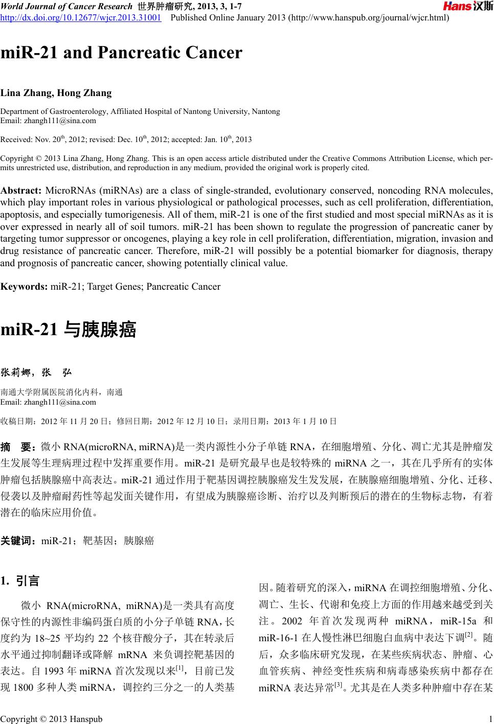
 World Journal of Cancer Research 世界肿瘤研究, 2013, 3, 1-7 http://dx.doi.org/10.12677/wjcr.2013.31001 Published Online January 2013 (http://www.hanspub.org/journal/wjcr.html) miR-21 and Pancreatic Cancer Lina Zhang, Hong Zhang Department of Gastroenterology, Affiliated Hospital of Nantong University, Nantong Email: zhangh111@sina.com Received: Nov. 20th, 2012; revised: Dec. 10th, 2012; accepted: Jan. 10th, 2013 Copyright © 2013 Lina Zhang, Hong Zhang. This is an open access article distributed under the Creative Commons Attribution License, which per- mits unrestricted use, distribution, and reproduction in any medium, provided the original work is properly cited. Abstract: MicroRNAs (miRNAs) are a class of single-stranded, evolutionary conserved, noncoding RNA molecules, which play important roles in various physiological or pathological processes, such as cell proliferation, differentiation, apoptosis, and especially tumorigenesis. All of them, miR-21 is one of the first studied and most special miRNAs as it is over expressed in nearly all of soil tumors. miR-21 has been shown to regulate the progression of pancreatic caner by targeting tumor suppressor or oncogenes, playing a key role in cell proliferation, differentiation, migration, invasion and drug resistance of pancreatic cancer. Therefore, miR-21 will possibly be a potential biomarker for diagnosis, therapy and prognosis of pancreatic cancer, showing potentially clinical value. Keywords: miR-21; Target Genes; Pancreatic Cancer miR-21 与胰腺癌 张莉娜,张 弘 南通大学附属医院消化内科,南通 Email: zhangh111@sina.com 收稿日期:2012 年11 月20 日;修回日期:2012 年12 月10日;录用日期:2013 年1月10日 摘 要:微小 RNA(microRNA, miRNA)是一类内源性小分子单链RNA,在细胞增殖、分化、凋亡尤其是肿瘤发 生发展等生理病理过程中发挥重要作用。miR-21 是研究最早也是较特殊的 miRNA 之一,其在几乎所有的实体 肿瘤包括胰腺癌中高表达。miR-21 通过作用于靶基因调控胰腺癌发生发发展,在胰腺癌细胞增殖、分化、迁移、 侵袭以及肿瘤耐药性等起发面关键作用,有望成为胰腺癌诊断、治疗以及判断预后的潜在的生物标志物,有着 潜在的临床应用价值。 关键词:mi R- 2 1 ;靶基因;胰腺癌 1. 引言 微小 RNA(microRNA, miRNA)是一类具有高度 保守性的内源性非编码蛋白质的小分子单链RNA,长 度约为 18~25 平均约 22 个核苷酸分子,其在转录后 水平通过抑制翻译或降解 mRNA 来负调控靶基因的 表达。自1993 年miRNA 首次发现以来[1],目前已发 现1800 多种人类miRN A ,调控约三分之一的人类基 因。随着研究的深入,miRNA 在调控细胞增殖、分化、 凋亡、生长、代谢和免疫上方面的作用越来越受到关 注。2002 年首次发现两种 miRNA ,miR-15a 和 miR-16-1 在人慢性淋巴细胞白血病中表达下调[2]。随 后,众多临床研究发现,在某些疾病状态、肿瘤、心 血管疾病、神经变性疾病和病毒感染疾病中都存在 miRNA 表达异常[3]。尤其是在人类多种肿瘤中存在某 Copyright © 2013 Hanspub 1 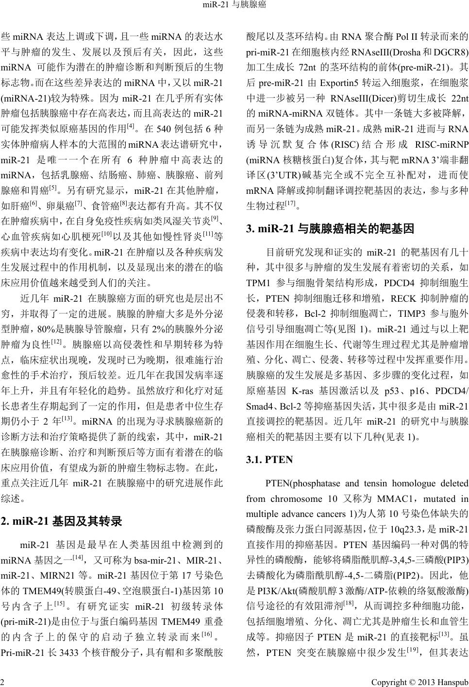 miR-21 与胰腺癌 些miRNA 表达上调或下调,且一些miRNA 的表达水 平与肿瘤的发生、发展以及预后有关,因此,这些 miRNA 可能作为潜在的肿瘤诊断和判断预后的生物 标志物。而在这些差异表达的miRNA 中,又以 miR-21 (miRNA-21)较为特殊。因为miR-21 在几乎所有实体 肿瘤包括胰腺癌中存在高表达,而且高表达的miR- 2 1 可能发挥类似原癌基因的作用[4]。在540 例包括 6种 实体肿瘤病人样本的大范围的 miRNA 表达谱研究中, miR-21 是唯一一个在所有 6种肿瘤中高表达的 miRNA,包括乳腺癌、结肠癌、肺癌、胰腺癌、前列 腺癌和胃癌[5]。另有研究显示,miR-21 在其他肿瘤, 如肝癌[6]、卵巢 癌 [7]、食管癌[8]表达都有升高。其不仅 在肿瘤疾病中,在自身免疫性疾病如类风湿关节炎[9]、 心血管疾病如心肌梗死[10]以及其他如慢性肾炎[11]等 疾病中表达均有变化。miR-21 在肿瘤以及各种疾病发 生发展过程中的作用机制,以及显现出来的潜在的临 床应用价值越来越受到人们的关注。 近几年 miR-21 在胰腺癌方面的研究也是层出不 穷,并取得了一定的进展。胰腺的肿瘤大多是外分泌 型肿瘤,80%是胰腺导管腺瘤,只有 2%的胰腺外分泌 肿瘤为良性[12] 。胰腺癌以高侵袭性和早期转移为特 点,临床症状出现晚,发现时已为晚期,很难施行治 愈性的手术治疗,预后较差。近几年在我国发病率逐 年上升,并且有年轻化的趋势。虽然放疗和化疗对延 长患者生存期起到了一定的作用,但是患者中位生存 期仍小于 2年[13]。miRNA 的出现为寻求胰腺癌新的 诊断方法和治疗策略提供了新的线索,其中,miR- 2 1 在胰腺癌诊断、治疗和判断预后等方面有着潜在的临 床应用价值,有望成为新的肿瘤生物标志物。在此, 重点关注近几年 miR-21 在胰腺癌中的研究进展作此 综述。 2. miR-21 基因及其转录 miR-21 基因是最早在人类基因组中检测到的 miRNA 基因之一[14],又可称为 bsa-mir-21、MIR-21、 miR-21、MIRN21 等。miR-21 基因位于第 17 号染色 体的 TMEM49(转膜蛋白-49、空泡膜蛋白-1)基因第 10 号内含子上[15] 。有研究证实 miR-21 初级转录体 (pri-miR-21)是由位于与蛋白编码基因 TMEM49 重叠 的内含子上的保守的启动子独立转录而来[16] 。 Pri-miR-21长3433 个核苷酸分子,具有帽和多聚酰胺 酸尾以及茎环结构。由RNA聚合酶 Pol II 转录而来的 pri-miR-21 在细胞核内经 RNAseIII(Drosha 和DGCR8) 加工生成长72nt 的茎环结构的前体(pre-miR-21)。其 后pre-miR-21 由Exportin5 转运入细胞浆,在细胞浆 中进一步被另一种 RNAseIII(Dicer)剪切生成长 22nt 的miRNA-miRNA 双链体。其中一条链大多被降解, 而另一条链为成熟miR- 2 1 。成熟miR-21 进而与RNA 诱导沉默复合体(RISC) 结合形成 RISC-miRNP (miRNA 核糖核蛋白)复合体,其与靶 mRNA 3’端非翻 译区(3’UTR)碱基完全或不完全互补配对,进而使 mRNA 降解或抑制翻译调控靶基因的表达,参与多种 生物过程[17]。 3. miR-21 与胰腺癌相关的靶基因 目前研究发现和证实的 miR-21 的靶基因有几十 种,其中很多与肿瘤的发生发展有着密切的关系,如 TPM1 参与细胞骨架结构形成,PDCD4 抑制细胞生 长,PTEN 抑制细胞迁移和增殖,RECK 抑制肿瘤的 侵袭和转移,Bcl-2 抑制细胞凋亡,TIMP3 参与胞外 信号引导细胞凋亡等(见图1)。miR-21 通过与以上靶 基因作用在细胞生长、代谢等生理过程尤其是肿瘤增 殖、分化、凋亡、侵袭、转移等过程中发挥重要作用。 胰腺癌的发生发展是多基因、多步骤的变化过程,如 原癌基因 K-ras 基因激活以及p53、p16、PDCD4/ Smad4、Bcl-2 等抑癌基因失活,其中很多是由 miR- 2 1 直接调控的靶基因。近几年 miR-21 的研究中与胰腺 癌相关的靶基因主要有以下几种(见表 1)。 3.1. PTEN PTEN(phosphatase and tensin homologue deleted from chromosome 10 又称为MMAC1,mutated in multiple advance cancers 1)为人第 10号染色体缺失的 磷酸酶及张力蛋白同源基因,位于10q 23.3,是 miR-2 1 直接作用的抑癌基因。PTEN 基因编码一种对偶的特 异性的磷酸酶,能够将磷脂酰肌醇-3,4,5-三磷酸(PIP3) 去磷酸化为磷脂酰肌醇-4,5-二磷脂(PIP2)。因此,他 是PI3K/Akt(磷酸肌醇 3激酶/ATP-依赖的络氨酸激酶) 信号途径的有效阻滞剂[18],从而调控多种细胞功能, 包括细胞增殖、分化、凋亡尤其是肿瘤生长和血管生 成等。抑癌因子 PTEN 是miR-21的直接靶标[13]。虽 然,PTEN 突变在胰腺癌中很少发生[19],但其表达 Copyright © 2013 Hanspub 2  miR-21 与胰腺癌 Copyright © 2013 Hanspub 3 Figure 1. miR-21 activity in cancer cells[16] 图1. miR-21在肿瘤细胞中的作用机制示意图[16] Table 1. The pancreatic cancer related target gene of miR-21 表1. miR-21与胰腺癌相关的靶基因 靶基因 参与的信号途径 作用 参考文献 PTEN PI3K/Akt/VEGF 促进细胞凋亡,抑制肿瘤生长和血管生成 [17-21] PDCD4 MAPK 促进细胞凋亡 [23-26] TIMP3 MMPS 抑制肿瘤生长、血管生成和侵袭 [28,29] Bcl-2 细胞凋亡信号途径 抑制细胞凋亡 [30,34] 减少可能为癌变的胰腺细胞提供有利的生长因素[20]。 M. Krausch[21]等人利用免疫组化检测了 38 例胰腺内 分泌肿瘤病人肿瘤组织中PTEN的表达并分析了 PTEN 表达与临床和病理的各项参数如性别、年龄、 TNM 分期等的相关性,发现免疫组化PTEN 染色阴 性的患者的生存率要明显优于阳性的患者,且 PTEN 失表达与 TNM分期正相关。另有研究显示 PTEN 和 Smad4 协同作用抑制小鼠胰腺导管腺癌的形成[22]。 3.2. PDCD4 PDCD4(programmed cell death 4)基因是 miR-21 直接作用的抑癌基因,位于人第十号染色体 10q24。 PDCD4 蛋白在包括造血干细胞、衰老的成纤维细胞以 及正常的肠和乳腺导管上皮细胞等多种细胞中均有 表达,主要分布于细胞核,介导多种生物过程,如细 胞周期进程、细胞存活等[23]。PDCD4 蛋白通过与翻 译启动子 RNA和ATP 依赖性 RNA 解旋酶——eIF4A 和eIF4G 相互作用靶向调控翻译过程。并且下调 PDCD4 表达可引起激活物蛋白-1(AP-1)依赖性转录、 actine/Tcf 依赖性转录和有丝分裂素蛋白激酶激酶类 1 的激活,并引起基质金属蛋白酶组织抑制因子的减 少,从而抑制肿瘤发生、发展、增殖、侵袭和转移[24,25]。 PDCD4 是miR-21 直接作用的抑癌基因,研究表明下 调miR-21 的表达可引起 PDCD4 表达上调。Imran Bhatti[26]等研究发现抑制胰腺癌细胞株 MI A-Pa-Ca-2 中miR-21 的表达可引起细胞增殖减少和细胞凋亡增 加,并同时伴随抑癌基因 PDCD4表达水平的增高, 提示 miR-21 可以作为上调 PDCD4 表达以治疗胰腺癌 的可能的治疗靶点。 3.3. TIMP3 TIMP3(tissue inhibitor of metalloproteinase)属于  miR-21 与胰腺癌 TIMPs 家族,此家族包含 4个成员,特异性地抑制基 质金属蛋白酶的蛋白水解活性[27]。与其他家族成员不 同的是,TIMP3 编码分泌的 24kDa 的蛋白结合于细胞 外基质。TIMP3 也是抑癌基因,其编码的蛋白能抑制 肿瘤生长、血管生成和侵袭,并促进细胞凋亡[28]。胰 腺癌存在大量TIMP3 的减少或缺失,尽管其启动子甲 基化的发生率较低[29]。目前研究发现,在胰腺癌中 miR-21 高表达与 TIMP3下调相关。因此,miR-21 诱 导的 TIMP3 下调很可能是又一个胰腺癌变的机制。 Yuichi Nagao[30]等人通过免疫组化分析了 65 例胰腺癌 病人和 5例正常人胰腺组织中PDCD4 和TIMP3 的表 达,发现与正常胰腺组织相比胰腺癌 PDCD4 和 TIMP3 的表达明显减少,数据显示 miR-21 高表达通 过下调抑癌基因 PDCD4和TIMP3的表达影响胰腺导 管腺癌的生物学行为。 3.4. Bcl-2 Bcl-2(B-cell lymphoma 2)属于 Bcl-2基因家族,主 要生物学功能是延长细胞寿命,增加细胞对各种凋亡 刺激因素的抵抗力,从而抑制凋亡。Bcl-2 基因家族 是研究最早的与细胞凋亡相关的基因,也是目前最受 关注的调控细胞凋亡的基因基因家族。Bcl-2 家族包 括凋亡抑制基因(Bcl-2,Bcl-xl,Bcl-w,Bfl-1,Brag-1, Mcl-1 和A1)以及促凋亡基因(Bax,Bak,Bcl-xs,Bad, Bik 和Hrk)[31]。细胞是否对凋亡信号有反应取决于凋 亡抑制因子和促凋亡因子的比例[32]。而 miR-21 是否 直接靶向作用于 bcl-2 仍然存在争议,有学者认为一 些miRNA 可以上调某些基因或蛋白的表达,但通常 不是直接作用[33,34]。但也有研究显示miR-21 是直接 作用于 bcl-2,参与调控肿瘤细胞凋亡。Jie Dong[35]等 人将 miR-21 类似物转染入 MIA-PaCa-2 胰腺癌细胞株 后,Bcl-2 表达明显上调而 Bax表达下调;相反将 miR-21 抑制剂转染入胰腺癌细胞株后出现了与以上 相反的结果。所有结果证实miR-21 可以上调Bcl-2 的 表达而下调 Bax 的表达;并构建具有 Bcl-2 mRNA 3’UTR 野生型和突变型的miR-21 结合位点的质粒转 染入细胞,采用荧光素酶报告监测发现与突变型的细 胞相比,野生型的细胞荧光素酶的活性在转染入 miR-21 拟似物后显著增加,相反在转染入 miR-21 抑制剂后显著减少,从而指出 miR-21 通过直接靶向 Bcl-2 mRNA 3’UTR来上调 Bcl-2 的表达。另外,抑 制miR-21表达可以上调乳腺癌[36]、胰腺癌细胞株中 Bcl-2 的表达而下调胶质母细胞瘤[37]中Bcl-2 的表达。 这提示 miR-21 对Bcl-2 表达的影响可能具有细胞类 型特异性。 4. miR-21 与胰腺癌的诊断、治疗以及预后 的关系 临床研究中,首次在人胶 质母细胞瘤中发 现 miR-21 高表达[38]。因此,miR-21 的异常表达可能是 肿瘤的一般特性,可以作为潜在的生物标志物,显现 出很高的临床应用价值。由于胰腺癌恶性程度高,易 于早期转移,难于早期诊断,预后较差,发病率逐年 上升,一直以来都是困扰医学研究者的难题。miRNA 的研究为胰腺癌研究提供了新的线索,尤其是 miR-2 1 在对胰腺癌诊断、治疗以及预后等方面的作用更是进 行了大量的研究。 4.1. miR-21 与胰腺癌的诊断 Kong[39]等用实时定量 PCR 检测了 35例胰腺导管 腺癌、15例慢性胰腺炎和 15 例正常人血清 miRN A 表达情况,结果显示血清 miR-21 可以区别胰腺癌患 者和正常人。由于 miR-21 在几乎所有的实体肿瘤中 都高表达,因此单独应用 miR-21 作为胰腺癌诊断的 标志物缺乏特异性。但有研究显示 miR-21 的表达水 平与胰腺癌术前血清 CA-199 的水平直接相关[40]。联 合传统的胰腺癌诊断标志物CA-199 可以提高胰腺癌 早期诊断的敏感性和特异性。目前,很多学者都致力 于寻找可应用与早期诊断的胰腺癌特异性的 miRNA 表达谱,miR-21 同样是其中不可缺少的miRNA. Rui Liu[41]等人收集 197 例胰腺癌病人的血清样本和 158 例正常人血清样本作为对照,筛选出 7种在胰腺癌中 差异表达的,其中包括miR - 21 。7种miRNA 组成的 表达谱作为胰腺癌诊断标志物在区别不同阶段胰腺 癌病人和非癌症人上具有高度敏感性和特异性,并且 能够正确区分胰腺癌和慢性胰腺炎的病人。 4.2. miR-21 与胰腺癌的治疗 胰腺癌除了外科手术治疗,化疗也是临床常用的 主要治疗手段。因此,预测化疗反应尤为重要。近期 Copyright © 2013 Hanspub 4  miR-21 与胰腺癌 众多研究显示 miRNA 在胰腺癌抗药性方面起一定的 作用。研究证实 miR-21 在胰腺癌细胞中能够诱导对 化疗药吉西他滨的化疗抵抗,miR-21 可以作为胰腺癌 化疗抵抗的生物标志物。另有研究尝试通过降低 miR-21 的表达来调控胰腺癌对吉西他滨的药物抵抗 ——吉西他滨抵抗的胰腺癌细胞经姜黄素类似物处理 以后 miR-21 表达下调,从而提高了吉西他滨的作用 敏感性[42];Jong-Kook Park[43]等研究发现使用反义寡 聚核苷酸来抑制 miR-21 的表达可减少胰腺癌细胞增 殖、阻滞细胞周期、诱导肿瘤细胞凋亡,并且可增强 吉西他滨对胰腺癌的作用敏感性。另外为了探讨 5-氟 尿嘧啶(5-FU)抗药性的机制以及miR-21 在抗药性方 面的作用,有研究反义抑制药物敏感细胞和抗药性细 胞中 miR-21 的表达从而引起 5-FU 活性的增加[44]。 miR-21 前体转染可显著降低抗肿瘤药物的药效并且 显著减低胞苷所诱导的细胞凋亡,而基质金蛋白酶 MMP-2/MMP-9 及血管内皮生长因子的表达明显上 调。添加肌醇磷脂 3激酶和抗真菌类抗生素后可减低 磷酸化 AKt 的水平,显著改善 miR-21 前体所诱导的 对抗癌药物的抗药性[45]。影响肿瘤细胞侵袭及转移的 因素如细胞凋亡程序的调整,蛋白激酶的磷酸化,基 因表达的改变共同参与了 miR-21 在胞苷抗药性中的 作用,这些发现为胰腺癌靶向联合治疗提供了新的方 法。 4.3. miR-21 与胰腺癌的预后 miR-21 是目前研究较多的可能与胰腺癌预后密 切相关的miR N A。无论在转移的癌组织还是癌旁组织 中,MiR-21 高表达的胰腺癌病人的总体生存率明显降 低,说明 miR-21 高表达与预后差密切相关[44,45]。 Dillhoff[46]等在胰腺癌研究中用原位杂交的方法证实 miR-21 的表达与肿瘤大小、分化、淋巴结转移及 T 分期无关,但是在无淋巴结肿大的亚组分析中, miR-21 高表达则提示预后较差。Ioannis[47] 等采用 RT-PCR检测了 88 例胰腺导管腺癌病人及 98例正常 对照的胰腺组织样中 miR-2 1,miR- 3 1,miR-122, miR-145,miR- 155,miR-146a,miR-210,and miR-222 的表达,经统计分析发现miR-21和miR-155 与肿瘤 分期和预后差密切相关,并且存在高表达组的胰腺癌 病人总体生存率显著低于低表达组,并指出 miR- 2 1 和miR-155 可以作为判断预后的独立因素。 5. miR-21 在胰腺癌研究中存在问题 虽然近几年 miR-21 在胰腺癌研究方面取得了一 定的进展,但总体来看尚处于初步阶段。 一方面,miR-21 靶基因众多。几项研究运用计算 法预测 miR-21 的可能靶 mRNA 有数百种[48],其中通 过实验研究证实的仅占少数。一种靶基因可受多种 miRNA 调控,而一种miRNA 又可以调控多种靶基因, 因此就形成了复杂的调控网络。并且在不同的细胞环 境中,一种 miRNA可能依赖一组特定的有效的靶 mRNA 调控不同的信号通路,从而引起不同的表型[49]。 这就带来了以下几个问题:一是实验技术方法上如何 简单有效的识别 miR-21 在胰腺癌发生发展中起关键 作用的特异性的靶基因。二是给探讨其在肿瘤发生发 展过程中的作用机制带来了很大的困难。因为 miRNA 可以调控 mRNA 稳定性及其翻译生产蛋白质,因此 miR-21 靶基因可以通过其 mRNA 和表达受 miR-21 影 响的蛋白质来识别。目前多采用 mRNA 表达分析和蛋 白组学的方法来识别 miR-21 的靶基因,两种方法各 有利弊。 另一方面,miR-21 在众多疾病包括肿瘤、心血管 疾病、免疫系统疾病等发生发展过程中都存在异常表 达,而且在众多肿瘤包括胰腺癌中表达上调。单独作 为胰腺癌诊断标志物明显缺乏特异性,且没有统一的 判断标准,但是联合其他诊断方法或寻找特异性的 miRNA 表达谱将可能成为今后肿瘤诊断标志物研究 方面的新方向,可以作为潜在的临床诊断标志物。 miR-21 在胰腺癌治疗和判断预后方面具有潜在的临 床应用价值,但仍处于初步阶段,还不能广泛应用于 临床。 6. 结语 越来越多的研究显示 miRNA 与肿瘤的发生、发 展有着密切的关系,在肿瘤增殖、侵袭和转移中起着 重要作用。miR-21 是在多种疾病包括肿瘤中起关键作 用的一类miR N A。在胰腺癌中 miR-21 表达上调并伴 随其靶基因的相应变化,这不仅为探索胰腺癌的发生 发展机制提供新的研究线索,也为胰腺癌诊断、治疗 以及判断预后方面提供好的临床思路。 Copyright © 2013 Hanspub 5  miR-21 与胰腺癌 参考文献 (References) [1] R. C. Lee, R. L. Feinbaum and V. Ambros. The C. elegans het- erochronic gene lin-4 encodes small RNAs with antisense com- plementarity to lin-14. Cell, 1993, 75: 843-854. [2] G. A. Calin, C. D. Dumitru, M. Shimizu, et al. Frequent deletions and down-regulation of micro-RNA genes miR15 and miR16 at 13q14 in chronic lymphocytic leukemia. Proceedings of the Na- tional Academy of Sciences of the USA, 2002, 99: 15524-15529. [3] Z. Liu, A. Sall and D. Yang. MicroRNA: An emerging therapeu- tic target and intervention tool. International Journal of Molecu- lar Sciences, 2008, 9: 978-999. [4] X. Fu, Y. Han, Y. Wu, et al. Prognostic role of microRNA-21 in various carcinomas: A systematic review and meta-analysis. Euro- pean Journal of Clinical Investigation, 2011, 41(11): 1245-1253. [5] S. Volinia, G. A. Calin, C. G. Liu, et al. A microRNA expression signature of human solid tumors defines cancer gene targets. Proceedings of the National Academy of Sciences of the USA, 2006, 103: 2257-2261. [6] Y. Tomimaru, H. Eguchi, H. Nagano, et al. Circulating micro- RNA-21 as a novel biomarker for hepatocellular carcinoma. Jour- nal of Hepatology, 2012, 56(1): 167-175. [7] K. E. Resnick, H. Alder, J. P. Hagan, et al. The detection of dif- ferentioally expressed microRNAs from the serum of ovarian cancer patients using a novel real-time PCR platform. Gyneco- logic Oncology, 2009, 112(1): 55-59. [8] S. Komatsu, D. Ichikawa, H. Takeshita, et al. Circulating mi- croRNAs in plasma of patients with oesophageal squmaous cell carcinoma. British Journal of Cancer, 2011, 105(1): 104-111. [9] H. Wang, W. Peng, X. Ouyang, et al. Circulating microRNAs as candidate biomarkers in patients with systemic lupus erythema- tosus. Translational Research, 2012, 160(3): 198-206. [10] T. Thum, C. Gross, J. Fiedler, et al. MicroRNA-21 contributes to myocardial disease by stimulating MAP kinase signalling in fi- broblasts. Nature, 2008, 456(7224): 980-984. [11] C. S. Neal, M. Z. Michael, L. K. Pimlott, et al. Circulating mi- croRNA expression is reduced in chronic kidney disease. Ne- phrology Dialysis Transplantation, 2011, 26(11): 3794-3802. [12] I. G. Papaconstantinou, A. Manta, M. Gazouli, et al. Expression of MicroRNAs in patients with pancreatic cancer and its prog- nostic significance. Pancreas, 2013, 42(1): 67-71. [13] C.-K. Yu, S.-N. Yu, Z.-H. Lu, et al. Research advances in mi- croRNA in pancreatic ductal adenocarcinoma. Acta Academiae Medicinae Sinicae, 2011, 33(5): 575-581. [14] X. Cai, C. H. Hagedorn and B. R. Cullen. Human microRNAs are processed from capped, polyadenylated transcripts that can also function as mRNAs. RNA, 2004, 10(12): 1957-1966. [15] B. Qian, D. Katsaros, L. Lu, M. Preti, A. Durando, R. Arisio, L. Mu and H. Yu. High miR-21 expression in breast cancer associ- ated with poor disease-free survival in early stage disease and high TGF-β1. Breast Cancer Research and Treatment, 2009, 117 (1): 131-140. [16] S. Fujita, T. Ito, T. Mizutani, S. Minoguchi, N. Yamamichi, K. Sakurai and H. Iba. MiR-21 gene expression triggered by AP-1 is sustained through a double-negative feedback mechanism. Journal of Molecular Biology, 2008, 378: 492-504. [17] S. D. Selcuklu, M. T. Donoghue and C. Spillane. MiR-21 as a key regulator of oncogenic processes. Biochemical Society Trans- actions, 2009, 37: 918-925. [18] M. Krausch, A. Raffel, M. Anlauf, et al. Loss of PTEN expres- sion in neuroendocrine pancreatic tumors. Hormone and Meta- bolic Research, 2011, 43: 865-871. [19] A. Sakurada, A. Suzuki, M. Sato, H. Yamakawa, et al. Infrequent genetic alterations of the PTEN/MMAC1 gene in Japanese pa- tients with primary cancers of the breast, lung, pancreas, kidney, and ovary. Japanese Journal of Cancer Research, 1997, 88: 1025- 1028. [20] M. Escriva, S. Peiro, N. Herranz, et al. Repression of PTEN phosphatase by snail1 transcriptional factor during gamma ra- diation-induced apoptosis. Molecular and Cellular Biology, 2008, 28: 1528-1540. [21] M. Krausch, A. Raff el, M. Anlauf, et al. Loss of PTEN expres- sion in neuroendocrine pancreatic tumors. Hormone and Meta- bolic Research, 2011, 43: 865-871. [22] X. Xu, B. Ehdaie, N. Ohara, et al. Synergistic action of Smad4 and PTEN in suppressing pancreatic ductal adenocarcinoma for- mation in mice. Oncogene, 2010, 29: 674-686. [23] B. Lankat-Buttgereit, R. Goke. The tumour suppressor Pdcd4: Recent advances in the elucidation of function and regulation. Biology of the Cell, 2009, 101: 309-317. [24] R. Nieves-Alicea, N. H. Colburn, A. M. Simeone, et al. Pro- grammed cell death 4 inhibits breast cancer cell invasion by in- creasing tissue inhibitor of metalloproteinases-2 expression. Breast Cancer Research and Treatment, 2009, 114: 203-209. [25] Z. T. Wei, X. Zhang, X. Y. Wang, et al. PDCD4 inhibits the ma- lignnant phenotype of ovarian cancer cells. Cancer Science, 2009, 100: 1408-1413. [26] I. Bhatti, A. Lee, V. James, et al. Knockdown of microRNA-21 inhibits proliferation and increases cell death by targeting pro- grammed cell death 4 (PDCD4) in pancreatic ductal adenocar- cinoma. Journal of Gastrointestinal Surgery, 2011, 15: 199-208. [27] L. Blavier, P. Henriet, S. Imren, et al. Tissue inhibitors of matrix metalloproteinases in cancer. Annals of the New York Academy of Sciences, 1999, 878: 108-119. [28] M. Ahonen, M. Poukkula, A. H. Baker, et al. Tissue inhibitor of metalloproteinases-3 induces apoptosis in melanoma cells by stabilization of death receptors. Oncogene, 2003, 22: 2121-2134. [29] V. Fendrich, E. P. Slater, E. Heinmoller, et al. Alterations of the tissue inhibitor of metalloproteinase-3 (TIMP3) gene in pancre- atic adenocarcinomas. Pancreas, 2005, 30: e40-e45. [30] Y. Nagao, M. Hisaoka, A. Matsuyama, et al. Association of mi- croRNA-21 expression with its targets, PDCD4 and TIMP3, in pancreatic ductal adenocarcinoma. Modern Pathology, 2012, 25 (1):112-121. [31] R. J. Youle, A. Strasser. The BCL-2 protein family opposing activities that mediate cell death. Nature Reviews Molecular Cell Biology, 2008, 9: 47-59. [32] D. Chen, X. F. Zheng, D. L. Kang, et al. Apoptosis and expres- sion of the Bcl-2 family of proteins and P53 in human pancreatic ductal adenocarcinoma. Medical Principles and Practice, 2012, 21: 68-73. [33] F. Meng, R. Henson, H. Wehbe-Janek, et al. MicroRNA-21 re- gulates expression of the PTEN tumor suppressor gene in human hepatocellularcancer. Gastroenterology, 2007, 133: 647-658. [34] X. Zhou, Y. Ren, L. Moore, et al. Downregulation of miR-21 in- hibits EGFR pathway and suppresses the growth of human glio- blastoma cells independent of PTEN status. Laboratory Investi- gation, 2010, 90(2): 144-155. [35] J. Dong, Y. P. Zhao, L. Zhou, et al. Bcl-2 upregulation induced by miR-21 via a direct interaction is associated with apoptosis and chemoresistance in MIA PaCa-2 pancreatic cancer cells. Archives of Medical Research, 2011, 42: 8-14. [36] N. S. Wickramasinghe, T. T. Manavalan, S. M. Dougherty, et al. Estradiol downregulates miR-21 expression and increases miR- 21 target gene expression in MCF-7 breast cancer cells. Nucleic Acids Research, 2009, 37: 2584-2595. [37] X. Zhou, Y. Ren, L. Moore, et al. Downregulation of miR-21 inhibits EGFR pathway and suppresses the growth of human glioblastoma cells independent of PTEN status. Laboratory In- vestigation, 2010, 90: 144-155. [38] J. A. Chan, A. M. Krichevsky and K. S. Kosik. MicroRNA-21 is an antiapoptotic factor in human glioblastoma cells. Cancer Re- search, 2005, 65: 6029-6033. [39] X. Kong, Y. Du, G. Wang, et al. Detection of differentially ex- pressed microRNAs in serum of pancreatic ductal adenocarci- noma patients: MiR-196a could be a potential marker for poor prognosis. Digestive Diseases and Sciences, 2010, 56(2): 602- 609. [40] F. Tavano, F. F. di Mola, A. Piepoli, et al. Changes in miR-143 and miR-21 expression and clinic opathologicalcorrelations in Copyright © 2013 Hanspub 6  miR-21 与胰腺癌 Copyright © 2013 Hanspub 7 pancreatic cancers. Pancreas, 2012, 41(8): 1280-1284. [41] R. Liu, X. Chen, Y. Q. Du, et al. Serum MicroRNA expression profile as a biomarker in the diagnosis and prognosis of pancre- atic cancer. Clinical Chemistry, 2012, 58: 3610-3618. [42] S. Ali, A. Ahmad, S. Banerjee, et al. Gemcitabine sensitivity can be induced in pancreatic cancer cells through modulation of miR-200 and miR-21 expression by curcumin or its analogue CDF. Cancer Research, 2010, 70: 3606-3617. [43] J. K. Park, E. J. Lee, C. Esau, et al. Antisense inhibition of mi- croRNA-21 or -221 arrests cell cycle, induces apoptosis, and sensitizes the effects of gemcitabine in pancreatic adenocarcino- ma. Pancreas, 2009, 38(7): e190-e199. [44] J. H. Hwang, J. Voortman, E. Giovannetti, et al. Identification of microRNA-21 as a biomarker for chemoresistance and clinical outcome following adjuvant therapy in resectable pancreatic cancer. PLoS One, 2010, 5(5): e10630. [45] E. Giovannetti, N. Funel, G. J. Peters, et al. MicroRNA-21 in pancreatic cancer: correlation with clinical outcome and phar- macologic aspects underlying its role in the modulation of gem- citabine activity. Cancer Research, 2010, 70(11): 4528-4538. [46] M. Dillhoff, J. Liu, Frankel W., et al. MicroRNA-21 is overex- pressed in pancreatic cancer and a potential predictor of survival. Journal of Gastrointestinal Surgery, 2008, 12(12): 2171-2176. [47] I. G. Papaconstantinou, A. Manta, M. Gazouli, et al. Expression of microRNAs in patients with pancreatic cancer and its prog- nostic significance. Pancreas, 2013, 42(1): 67-71. [48] A. Grimson, K. K. Farh, W. K. Johnston, et al. MicroRNA tar- geting specificity in mammals: Determinants beyond seed pair- ing. Molecular Cell, 2007, 27: 91-105. [49] A. M. Krichevsky, G. Gabriely. MiR-21: A small multi-faceted RNA. Journal of Cellular and Molecular Medicine, 2009, 13(1): 39-53. |