Asian Case Reports in Surgery
Vol.04 No.01(2015), Article ID:15595,5
pages
10.12677/ACRS.2015.41001
Clinical Research Progress of Pyogenic Granuloma after Burns
Hongliang Zhao1,2, Liankui Zhao2, Weihai Sun2, Gaosong Yang2, Lijing Jia3, Cuiping Zhang4
1Student in 2012 Doctor Class, Chinese Peoples Liberation Army General Hospital, Beijing
2Department of Burns and Plastic, The First Hospital of Shijiazhuang, Shijiazhuang Hebei
3Department of Endocrinology, The 3rd Hospital of Hebei Medical University, Shijiazhuang Hebei
4Wound Repair and Tissue Regeneration Laboratory, The First Affiliated Hospital, General Hospital of PLA, Beijing
Email: zcp666666@sohu.com
Received: Jun. 10th, 2015; accepted: Jun. 27th, 2015; published: Jun. 30th, 2015
Copyright © 2015 by authors and Hans Publishers Inc.
This work is licensed under the Creative Commons Attribution International License (CC BY).
http://creativecommons.org/licenses/by/4.0/


ABSTRACT
Pyogenic granuloma after burns is different from classic pyogenic granuloma for its clinical features and histological characteristics. Therapy plans of pyogenic granuloma are so different that outcomes of pyogenic granuloma are various. The etiology of pyogenic granuloma is still unknown. It is the benign lesion with the obvious proliferation of keratinocyte and capillary. The diagnosis of pyogenic granuloma is based on the case history, clinical features and the histological examination. Because the disease has a tendency to self heal and complications after the surgery. Conservative plan should be chose to treat cases with pyogenic granuloma after burns firstly.
Keywords:Burn, Pyogenic Granuloma, Surgery, Graft, Complication
烧伤后化脓性肉芽肿临床研究进展
赵洪良1,2,赵连魁2,孙卫海2,杨高松2,贾黎静3,张翠萍4
1解放军总医院2012级博士区队,北京
2石家庄市第一医院烧伤整形科,河北 石家庄
3河北医科大学第三医院内分泌科,河北 石家庄
4全军组织修复与再生实验室,解放军总医院第一附属医院,北京
Email: zcp666666@sohu.com
收稿日期:2015年6月10日;录用日期:2015年6月27日;发布日期:2015年6月30日

摘 要
烧伤后化脓性肉芽肿与经典的化脓性肉芽肿相比较具有独特的临床表现和组织学特点。目前烧伤后化脓性肉芽肿国内外诊治方法尚不规范,因此不同方法治疗效果差异很大。经过文献检索发现:烧伤后化脓性肉芽肿病因尚未明确,它是发生在II度烧伤后以角质细胞和毛细血管急性增生为主要组织学特点良性病变,其诊断依赖于病史、临床表现和组织学检查。因为该病有自愈倾向和手术后并发症两方面的原因,其治疗过程,宜首选保守治疗方案,必要时结合手术治疗。
关键词 :烧伤,化脓性肉芽肿,手术,植皮,并发症

1. 引言
化脓性肉芽肿,又称为毛细血管扩张性肉芽肿、小叶状毛细血管瘤,是一种常见疾病,多由于毛细血管和/或小静脉分叶状增生从而形成的息肉状良性肿瘤[1] [2] 。其病因尚未明确可能与创伤、感染、妊娠及生长因子等有关[3] -[6] 。其病理表现为大量毛细血管为特征的血管增生性肉芽肿组织,间质可见组织水肿、中性粒细胞和淋巴细胞浸润。该病多见于儿童,成人少见,临床表现主要以瘤样增生病变为主,单发多见,多发者少见,其表面光滑或有分叶,有蒂或无蒂。深红色或带黄白色小点,质脆弱易出血。国内外其临床治疗主要有保守治疗和全层手术切除治疗,而保守治疗,例如冷冻,激光,电灼存在复发的风险,所以全层切除在国内外被认为是疗效可靠的方法[1] [7] -[11] 。
烧伤后化脓性肉芽肿(pyogenic granuloma after burns, PGB)最早为阿根廷学者在1978年报道并被治愈,近年来国内外临床病例报道逐渐增多(表1),众多研究发现它既有毛细血管增生的病理特点,又有与经典的化脓性肉芽肿有许多不同的的独特临床表现[12] -[25] 。至今国内外对其临床诊治方法尚未规范,造成很多问题,其中最重要的是一些观点认为PGB应当首选手术治疗,而另一些观点认为该病可以经保守治疗后自愈,这矛盾的观点给临床实践带来很多困惑,为此笔者就PGB诊断和治疗等内容进展进行综述,希望对临床医生将来的诊治有所帮助。
2. 检索策略
英文主要资料库包括:PubMed、Cochrane图书馆、EMBASE和Web of science。检索策略:pyogenic granuloma and burns,语种为英语的文章,不包括综述。中文资料库包括:中文生物医学文献数据库、中国期刊全文数据库、中文科技期刊数据库、中国优秀博硕士学位论文全文数据库。检索策略:化脓性肉芽肿和烧伤,检索时间段至2014年12月,无发表类型限制。纳入标准:烧伤后化脓性肉芽肿患者,并有病理诊断结果。检索结果:中文:4篇,英文:10篇(表1)。
3. 结果
3.1. 烧伤后PGB病理机制
尽管PGB的病因尚未明确,但是II度烧伤作为首发因素起着关键作用。因为目前已经报道的31例
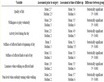
Table 1. 31 Cases of PGB were reported in general
表1. 已报道31例PGB病例一般资料
年龄:m为月,y为岁。性别:M为男性,F为女性。
患者中仅有1例患者PGB发生于烧伤后一年的瘢痕[23] ,其余30例患者PGB均发生于II度烧伤创面[12] -[22] [24] [25] 。II度烧伤创面非常常见,然而PGB患者罕见提示其他诱发因素的存在。已有研究发现多例热牛奶烧伤后出现PGB患者,因此认为牛奶中的某种物质可能导致PGB的发生[18] 。然而,另有研究认为PGB与牛奶的关系不大[13] 。近来研究表明微生物感染可能是PGB重要的促发因素,目前创面感染的细菌主要包括阴沟肠杆菌[16] 、表皮葡萄球菌和乙型链球菌[20] 和大肠杆菌[22] ,管电镜发现包涵体[16] ,但是病毒种类未明确。此外,细菌和真菌混合感染金黄色葡萄球菌绿脓杆菌和白色念珠菌也有报道[14] 。
值得注意的是,烧伤创面感染比较常见,但是为什么PGB只出现在II度烧伤创面是个值得考虑的问题。这可能与PGB的组织学特点有关。典型的PGB组织学改变包括以下内容:角质细胞过度增生,可见明显的上皮脚插入肿物深部,大量毛细血管增生,间质水肿并伴有炎性细胞浸润。与经典的化脓性肉芽肿相比较角质细胞的过度增生仅存在于烧伤后化脓性肉芽肿。由此可见,PGB发病可能是由于皮肤创面细菌感染后,不断释放内毒素或者其它物质,刺激组织局部产生大量细胞因子,这些细胞因子会促进皮肤创面各种修复细胞成分,如成纤维细胞、角朊细胞、血管内皮细胞等过度增殖[25] -[28] ,从而表现为局部角质细胞和真皮内毛细血管增生的组织特点。另一方面III度烧伤由于创面已经完全失去屏障,毛细血管的增生未受到周围组织的限制而形成典型的肉芽组织,而I度烧伤创面皮肤屏障结构尚完整有较强的抵抗能力,因此创面感染后足量的炎症因子或者生长因子的释放和基本完整的皮肤结构是PGB的出现重要因素,也就是说,当创面得到及时处理时,就不会出现PGB。
3.2. PGB诊断
3.2.1. 临床表现特点
文献报道发病年龄最小8个月,最大56岁,男女比例为21:10。烧伤原因依次为热液,火焰和电烧伤(见表1)。PGB发病部位包括头、面、颈部,四肢、躯干等部位均有报道,单发或者弥漫分布,大小不等,分布不均,病变主要局限在浅或深II度烧伤区域。该病发病急,出现时间为烧伤2~27天,病变最大者直径6 cm [19] 。
烧伤后PGB临床表现为发生在烧伤II度创面的急性增生和逐渐消退过程,这与经典化脓性肉芽肿罕见自愈特点明显不同。首先PGB大多数发生于II度烧伤创面,或者已经愈合烧伤创面,并局限在创面范围内,目前报道的仅有1例发生于烧伤后的瘢痕区域(表1)。临床表现为急性增生过程,而非慢性刺激过程,增殖局限在烧伤区域,在烧伤3 d ~ 12 d后局部烧伤区长出小米大小的红疹,并迅速长大,有的互相融合成片高出皮肤(图1),表面高低不平,色暗红,有的被覆一层上皮,表面有渗出物,破后流出血性渗出物,质较脆,易出血,创周炎症反应明显。经历增生高峰后逐渐消退的临床过程表现为肿物停止增大,并逐渐缩小。创面干燥,肿物创面周围炎症反应明显可以完全消退,恢复皮肤的正常结构。
3.2.2. 鉴别诊断
PGB的组织学特点提示其为良性病变的急性过程,因此容易与高分化鳞癌的组织学鉴别。例如根据病史烧伤后在II度烧伤创面发病,临床特点以及经病理学检查为良性病变等进行鉴别诊断。
3.2.3. 临床治疗应以保守治疗为首选
经典的化脓性肉芽肿烧伤治疗的方法较多,例如电灼、激光、刮除术、冷冻和手术,除了手术切除无复发外,其它方法都有一定的复发率。近年来,尽管不断的有新的方法报道[11] [29] -[31] ,但是复发的问题仍然不能避免。因此,目前国内外学者仍认为手术切术最可靠的方法[1] [7] 。
然而,因为PGB有自愈倾向和手术后并发症两方面的原因,其治疗原则宜首选保守治疗方案,必要时结合手术治疗。当前GB临床局部治疗总体上分为手术治疗和保守治疗。手术治疗中分为三类,PGB切除自体皮移植术或缝合术,未见刮除术应用于PGB的治疗。手术切除的适应症主要是对于临床治疗效果不明显的病例,PGB进行性增大的患者[14] [18] [19] [24] [25] 。尽管手术切除具有时间短,无复发的优点,但是有下列情况的患者不宜手术切除治疗:1) PGB位于重要部位,例如面部,颈部,以及重要关节部位。这是因为手术切除后遗留的瘢痕及色素异常对患者的形象和心理影响极大,以及手术后的瘢痕挛缩导致肢体运动障碍;2) 大面积弥散分布的婴幼儿患者。这是因为此类患者因在术中的出现大量出血,而需承担更大的手术风险。3) 正在逐渐消退的PGB患者也不宜承担麻醉的风险而进行手术治疗。

Figure 1. Large pyogenic granuloma after burns on a 4-year-old girl’s left leg buttock (left). Tissue section was stained with Hematoxylin-eosin (right), ×100
图1. 一个4岁女孩左大腿及臀部巨大烧伤后化脓性肉芽肿及苏木精一伊红染色切片结果,×100倍
保守治疗主要包括两方面的内容:一方面是创面的治疗,例如,清创术,选择适当的敷料保护创面。需要注意的是,根据PGB的上皮细胞和血管内皮细胞增殖的组织学特点,在这种创面应用表皮细胞生长因子使需要谨慎对待;另一方面是抗生素的应用,可根据创面或者活检组织细菌培养后的结果进行调整。既往研究认为经典化脓性肉芽肿病变深达皮下组织,电灼、冷冻和水平切除或刮除肿物时,因为以正常皮肤为参照皮肤平面瘤体保留偏多易导致复发,而切除平面如过深则不易愈合且有瘢痕增生或者需要植皮[1] [7] 。正是由于以上的问题,手术治疗经典化脓性肉芽肿一直受到临床医生的首选,但是对于PGB患者来说,目前报道患者中不但自愈的患者无复发现象,而且接受保守治疗的病例中也无复发出现。随着近年来报告病例逐渐增多,保守治疗近年来越来越受到重视,其主要原因:一个是术后并发症未见报道,另一个是避免了手术带给患者身心痛苦不能忽视。对于面部或者重要部位等多种情况不适合手术治疗的条件下,促使更多临床医生选择非手术治疗,并不断取得良好的效果。因此,依据烧伤后PGB临床表现,在病理检查确定为良性病变前提下,首选保守治疗,必要时结合手术治疗是一个谨慎治疗方案。
4. 展望
临床迫切需要烧伤后PGB相关研究以明确其病因及病理机制,指导临床医生选择最佳诊治方案,以减轻患者的痛苦,尤其是探索此类创面皮肤完全性再生过程中精细的调控过程的规律,对于深入推进伤口愈合以及上皮细胞—间充质细胞转化,皮肤肿瘤研究等具有巨大的和潜在的经济与社会效益。
资助信息
本研究受到课题资助国家自然基金项目(81121004,81230041,81171798,81100591)和国家973项目,2012CB518105)、国家科技重点项目(2011ZXJ07104B-03B),河北省医学科学研究重点课题计划20120171资助。
文章引用
赵洪良,赵连魁,孙卫海,杨高松,贾黎静,张翠萍, (2015) 烧伤后化脓性肉芽肿临床研究进展
Clinical Research Progress of Pyogenic Granuloma after Burns. 亚洲外科手术病例研究,01,1-6. doi: 10.12677/ACRS.2015.41001
参考文献 (References)
- 1. Greene, A.K. (2011) Management of hemangiomas and other vascular tumors. Clinics in Plastic Surgery, 38, 45-63.
- 2. 廖文俊, 高天文, 夏汝山, 赵小东, 李平, 刘玉峰 (2001) 烫伤后多发性化脓性肉芽肿的临床病理及电镜观察. 第四军医大学学报, 24, 2279-2281.
- 3. Harris, M.N., Desai, R., Chuang, T.Y., Hood, A.F. and Mirowski, G.W. (2000) Lobular capillary hemangiomas: An epidemiologic report, with emphasis on cutaneous lesions. Journal of the American Academy of Dermatology, 42, 1012-1016.
- 4. Lee, J. and Lynde, C. (2001) Pyogenic granuloma: pyogenic again? Association between pyogenic granuloma and Bartonella. Journal of Cutaneous Medicine and Surgery, 5, 467-470.
- 5. Tervahartiala, B. and Ainamo, J. (1989) The development during pregnancy of pyogenic granulomas superimposed upon a congertital hemaneioma: A case report. Journal of Periodontology, 60, 358-361.
- 6. Zhang, J.L., Wang, L., Jin, Y.C., Huang, Q.S., Jin, W.L. (2001) Analysis of VEGF and integrinβ3 mRNA expression during angiogenetic process in granulation tissue with RT-PCR. Journal of Nanjing Medical University, 2, 145-147.
- 7. Patrice, S.J., Wiss, K. and Mulliken, J.B. (1991) Pyogenic granuloma (lobbular capillary hemangioma): A clinicopathogic study of 178 cases. Pediatric Dermatology, 8, 267-276.
- 8. Tay, Y.K., Weston, W.L. and Morelli, J.G. (1997) Treatment of pyogenic granuloma in children with the flashlamp pumped pulsed dye laser. Pediatrics, 99, 368-370.
- 9. Kirschner, R.E. and Low, D.W. (1999) Treatment of pyogenic granuloma by shave excision and laser photocoagulation. Plastic and Reconstructive Surgery, 104, 1346-1349.
- 10. Mirshams, M., Daneshpazhooh, M., Mirshekari, A., Taheri, A., Mansoori, P. and Hekmat, S. (2006) Cryotherapy in the treatment of pyogenic granuloma. Journal of European Academy of Dermatology and Venereology, 20, 788-790.
- 11. 杨灿 (2011) 化脓性肉芽肿3种治疗方法比较. 中国临床医学, 3, 374-375.
- 12. De Kaminsky, A.R., Otero, A.C., Kaminsky, C.A., Shaw, M., Formentini, E. and Abulafia, J. (1978) Multiple disseminated pyogenic granuloma. British Journal of Dermatology, 98, 461-464.
- 13. Momeni, A.Z., Enshaieh, S., Sodifi, M. and Aminjawaheri, M. (1995) Multiple giant disseminated pyogenic granuloma in three patients burned by boiling milk. International Journal of Dermatology, 34, 707-710.
- 14. Ceyhan, M., Erdem, G., Kotiloğlu, E., Kale, G., Talim, B., Kanra, G. and Başaran, I. (1997) Pyogenic granuloma with multiple dissemination in a burn lesion. Pediatric Dermatology, 14, 213-215.
- 15. 侯松治, 李慧, 赵学兰, 彭馈英 (2005) 烧伤愈合创面继发化脓性肉芽肿样病变四例. 中华烧伤杂志, 1, 76.
- 16. Liao, W.J., Fan, P.S., Fu, M., Gao, T.W., Liu, Y.F. and Ikeda, S. (2006) Clinicopathological and ultrastructural study of multiple lobular capillary hemangioma after scalding. Dermatology, 213, 34-36.
- 17. Aliağaoğlu, C., Bakan, V., Atasoy, M. and Toker, S. (2006) Pyogenic granuloma with multiple and satellite involvement after a burn in a 5-year-old child. Journal of Dermatology, 2, 150-152.
- 18. Bozkurt, M., Külahçı, Y., Zor, F. and Aşkar, I. (2006) Multiple giant disseminated pyogenic granuloma in a burn lesion. Journal of Burn Care Research, 27, 247-249.
- 19. Diallo, M., Niang, S.O., Kane, A., Dieng, M. and Ndiaye, B. (2006) Pyogenic granulomas with multiple satellites spontaneously resolved. Nouvelles Dermatologiques, 25, 701-703.
- 20. 姚贵申, 苗国英, 王世君, 刘保国 (2007) 烫伤后多发性化脓性肉芽肿八例临床分析. 中华皮肤科杂志, 4, 237- 238.
- 21. Ceyhan, A.M., Basak, P.Y., Akkaya, V.B., Yildirim, M. and Kapucuoglu, N. (2007) A case of multiple, eruptive pyogenic granuloma developed on a region of the burned skin: Can erythromycin be a treatment option? Journal of Burn Care Research, 28, 754-757.
- 22. 印道春, 彭英霞 (2009) 烧伤后多发性化脓性肉芽肿1例. 中国皮肤性病学杂志, 12, 850.
- 23. Shirol, S.S., Nimbaragi, G., Choukimath, S.M. and Yenni, V.V. (2013) Lobular capillary hemangioma in a post-burn scar. European Journal of Plastic Surgery, 36, 323-326.
- 24. Durgun, M., Selçuk, C.T., Özalp, B., Aydinol, M. and Alabalik, U. (2013) Multiple disseminated pyo-genic granuloma after second degree scald burn: A rare two case. International Journal of Burns and Trauma, 3, 125-129.
- 25. Wiktorowska-Owczarek, A. (2013) The effect of valdecoxib on the production of growth factors evoked by hypoxia and bacterial lipopolysaccharide in HMEC-1 cells. Advances in Clinical and Experimental Medicine, 22, 795-800.
- 26. Mastrofrancesco, A., Kovacs, D., Sarra, M., Bastonini, E., Cardinali, G., Aspite, N., et al. (2014) Prec-linical studies of a specific PPARγ modulator in the control of skin inflammation. Journal of Investigative Dermatology, 134, 1001- 1011.
- 27. Vasconcelos, M.G., Alves, P.M., Vasconcelos, R.G., da Silveira, É.J.D., Medeiros, A.M. and de Queiroz, L.M.G. (2011) Expression of CD34 and CD105 as markers for angiogenesis in oral vascular malformations and pyogenic granulomas. European Archives of Oto-Rhino-Laryngology, 268, 1213-1217.
- 28. Yuan, K., Jin, Y.T. and Lin, M.T. (2000) The detection and comparison of angiogenesis-associated factors in pyogenic granuloma by immune-histochemistry. Journal of Periodontology, 71, 701-709.
- 29. Ghodsi, S.Z., Raziei, M., Tahefi, A., Karami, M., Mansoori, P. and Farnaghi, F. (2006) Comparison of cryotherapy and curettage for the treatment of pyogenic granuloma: A randomized trial. British Journal of Dermatology, 154, 671-675.
- 30. 吴亚芬, 李巍, 钱华, 鲁慧, 徐荣华 (2013) 儿童化脓性肉芽肿3种治疗方法疗效比较. 中国中西医结合皮肤性病学杂志, 4, 250-252.
- 31. Sud, A.R. and Tan, S.T. (2010) Pyogenic granuloma-treatment by shave-excision and/or pulsed-dye laser. Journal of Plastic, Reconstructive Aesthetic Surgery, 63, 1364-1368.