Biophysics
Vol.03 No.01(2015), Article ID:15078,5
pages
10.12677/BIPHY.2015.31001
Research Advances of AMMECR1
Huamin Zhou, Chengfeng Cai, Meng Xu, Guang Li
The State Key Laboratory of the Cellular Stress Biology, School of Life Sciences, Xiamen University, Xiamen Fujian
Email: huaminzhou@xmu.edu.cn
Received: Mar. 23rd, 2015; accepted: Apr. 8th, 2015; published: Apr. 15th, 2015
Copyright © 2015 by authors and Hans Publishers Inc.
This work is licensed under the Creative Commons Attribution International License (CC BY).
http://creativecommons.org/licenses/by/4.0/


ABSTRACT
AMMECR1 (Alport syndrome, mental retardation, midface hypoplasia, and elliptocytosis chromosomal region gene 1) is a gene from the novel X-linked contiguous gene deletion syndrome AMME critical region. It encodes a transcript that is conserved throughout the course of evolution. There is a considerable degree of homology between the AMMECR1 proteins from different species ranging from bacteria and archaea to eukaryotes. This conservation suggests that AMMECR1 and its homologue proteins may exert essential functions in a variety of organisms. In this review, we will describe that AMMECR1 expression, crystal structure, phosphorylation, function-related proteins, miRNAs targeting to AMMECR1 and its interaction partner to promote the study of AMMECR1.
Keywords:AMMECR1, Phosphorylation, Related Proteins, miRNA
AMMECR1的研究进展
周化民,蔡成峰,徐盟,李光
厦门大学生命科学学院,细胞应急生物学国家重点实验室,福建 厦门
Email: huaminzhou@xmu.edu.cn
收稿日期:2015年3月23日;录用日期:2015年4月8日;发布日期:2015年4月15日

摘 要
AMMECR1是X-连锁邻近基因缺陷综合症AMME关键区域基因之一。它是一个非常古老而且保守的基因,从古细菌,细菌,酵母、线虫、果蝇到哺乳类和人,这一蛋白都有很高的同源性,应该在基本的生物学过程中执行某个重要的功能。但AMMECR1的生物学功能研究还很匮乏。本文从AMMECR1的表达、同源蛋白的晶体结构、磷酸化、功能相关蛋白、靶向AMMECR1的miRNA、AMMECR1可能的相互作用蛋白等方面进行综述,为开展AMMECR1研究的提供参考。
关键词 :AMMECR1,磷酸化,相关蛋白,miRNA

1. 引言
AMMECR1 (Alport syndrome, mental retardation, midface hypoplasia, elliptosis chromosomal region gene 1)是X-连锁邻近基因缺陷综合症(contiguous gene deletion syndrome) AMME关键区域基因之一[1] 。AMMECR1是一个非常古老而且保守的基因,从古细菌,细菌,酵母、线虫、果蝇到哺乳类和人,这一蛋白都有很高的同源性,可以想见,它一定在细胞生命活动的核心过程中发挥作用。目前比较明确的是该区域内编码Ⅳ型胶原a-5链基因COL4A5点突变或基因内删除将引起血管性球性肾炎。然而带有不包含COL4A5基因但含有包括AMMECR1在内的9个基因的X染色体删除的患者,仍表现中等智力障碍(moderate intellectual disability),神经性听力损失(sensorineural hearing loss),面部发育不全(facial dysmorphism),幽门狭窄(pyloric stenosis)以及肠梗阻(intestinal obstruction) [2] 。不过,AMMECR1是否和这些病理现象直接相关有待进一步研究。
AMMECR1有6个外显子,产生4种拼接形式转录子(transcripts),最长的转录子编码333个氨基酸的多肽,是AMMECR1的常见形式;位居第二的转录子缺失了第二外显子(氨基酸159-195);如果翻译从mRNA 5′端第二个甲硫氨酸密码子开始则形成缺少N端123个氨基酸(氨基酸1-123)的多肽;第一外显子和其下游内含子中一个额外的外显子(外显子2′)拼接则形成仅含N端157个氨基酸的多肽[1] 。AMMECR1的生物学功能研究是极其匮乏的。本文探讨AMMECR1研究现状及可能参与的生物学过程。
2. AMMECR1的表达
本AMMECR1组织分布一般比较广泛,但还能在一些特殊细胞或条件下表达、不表达甚至被删除(表1)。如在兔白内障手术后水状液(aqueous humor,AH)样品中以及在养殖于含乙炔雌二醇(ethinylestradiol)水体达14天的鱼类精巢组织中均有表达[3] [4] ;但在鼠滋养层干细胞K4GFP中,AMMECR1不表达[5] ;而在智力障碍者的外周血白细胞及人类不同恶性程度的眼色素层黑色素瘤(Uveal melanoma)细胞,AMMECR1是染色体删除的 [2] [6] 。如果AMMECR1酵母(S. pombe)同源物Spac688.03c删除、酵母细胞对紫外线中等敏感 [7] ,因此,它可能和DNA的修复有关。
3. AMMECR1同源蛋白的晶体结构
Pyrococcus horikoshii OT3蛋白PH0010和人AMMECR1 C-端氨基酸序列类似, PH0010晶体结构显示它有大小两个结构域,两个结构域间有一个可变的裂隙 [8] ,结构比较分析显示,它含有一个RAGNYA (Ribosomal proteins L3 and L1, ATP grasp modules, the GYF domain, DNA recombination proteins of the NinB family from caudate bacteriophages, the C-terminal DNA-interacting domain of the Y-family DNA polymerases, the uncharacterized enzyme AMMECR1)折叠,像tRNA Wybutosine生物合成酶Tyw3p, AMMECR1可能催化某个RNA的碱基修饰 [9] 。
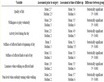
Table 1. AMMECR1 expression
表1. AMMECR1的表达
4. AMMECR1磷酸化
相对于其它AMMECR1同源物,人AMMECR1N端多出一段富含甘氨酸和丝氨酸的多肽。生物信息学分析显示AMMECR1有6个PKC (protein kinase C)磷酸化位点(S121、T150、T163、S249、T291、S299)和3个酪蛋白激酶(casein kinase II)磷酸化位点(T206、T221、S305),23个潜在的N-豆蔻酰化位点 [1] 。有趣的是,这些磷酸化位点都不在人AMMECR1富含甘氨酸和丝氨酸的N端,但质谱结果显示人AMMECR1有六个磷酸化位点(S15、S16、S19、S21、S29和S34) [10] - [13] ,它们都分布在富含甘氨酸和丝氨酸的N端,可见,需要实验证实人AMMECR1真正的磷酸化位点。
5. AMMECR1功能相关蛋白
AMMECR1可能参与多个细胞生物过程,受多个蛋白质或其他因子的调控(表2)。EPS8 (epidermal growth factor receptor pathway substrate 8,能增强细胞繁殖、迁徙及肿瘤发生)在HN4细胞(源自头颈部的原发性鳞状细胞癌细胞)中的过表达、转录因子TFAP2C(涉及乳腺发育、分化及肿瘤发生)在MCF7细胞中的过表达,均能上调AMMECR1的表达[14] [15] ;GBM (glioblastoma multiforme)是普通侵袭性原发性脑瘤,用葡萄多酚resveratrol (RV)刺激GBM-CD133+细胞,也能上调AMMECR1的表达[16] ;新生7天的小鼠在低氧(75%)中养5天,然后在正常空气中养12小时,视网膜AMMECR1的表达上调[17] 。PHF8 (plant homeodomain (PHD) finger-containing proteins 8,是组蛋白H3K9去甲基化酶)。在HeLa细胞中的低表达(knockdown),AMMECR1的表达下调[18] ;在IFN-γ/TNF-α联合刺激下,hcMSCs (human bone marrow (BM)-derived clonal mesenchymal stem cells (MSCs))细胞则能大幅下调AMMECR1的表达[19] ; FGFR4 (fibroblast growth factor receptor 4) 在HCC1.2. (hepatocellular carcinoma)中的表达及在高风险MDS (myelodysplastic syndrome)-CD34+细胞及红细胞样细胞(Erythroid cells)在添加BM-SCs (bone marrow stromal cells)细胞培养上清的培养基中培养,AMMECR1的表达均下调[20] -[22] 。在AML (acute myeloid leukemia)细胞,decitabine诱导MLL5 (human trithorax-group (Trx-G) gene)敲除的细胞和具有CBE (classic bladder exstrophy)的病人的膀胱组织,AMMECR1的表达显示明显差异[23] [24] 。这些蛋白或因子如何调控AMMECR1表达,目前还不清楚。AMMECR1表达变化显示可能参与在此种条件下细胞的某些生物学过程,如PHF8删除,导致X连锁智力发育迟缓,在HeLa细胞中,降低PHF8的表达,导致AMMECR1明显下调 [18] ,而在IFN-γ/TNF-α联合刺激下,hcMSCs细胞STAT2大幅上调,抑制T细胞繁殖,此时AMMECR1的表达大幅下调 [19] ,AMMECR1是否和智力发育及T细胞繁殖有关,这些或许值得探讨。
6. 靶向AMMECR1的miRNA
miRNAs (MicroRNAs)是真核生物中一类大小长约20~25个核苷酸、通常结合在mRNA的3′端非编码区的非编码RNA,miRNA参与各种各样的调节途径,包括发育、病毒防御、造血过程、器官形成、细胞增殖和凋亡、脂肪代谢等等生物信息学分析发现,很多miRNA可以靶向AMMECR1 (表3),如has-miR-517a、miR-191、has-miR-375和miR-124 [25] - [28] ;把T3BA细胞(hES (human embryonic stem)-T3 cells)培养在补充4 ng/ml bFGF (human basic fibroblast growth factor)和5 ng/ml activin A干细胞培养基中,miRNA let-7c表达仅占对照组的0.06,let-7c靶向基因AMMECR1表达升高15.68倍 [29] ;在MDA-MB-435乳腺癌细胞(不表达内源性integrinα6β4)中过表达integrinα6β4,miR-92ab和miR-99ab/100 family miRNA表达受抑,但它们的靶基因 AMMECR1表达小幅上扬 [30] ;表达miR-26b 也降低AMMECR1表达 [31] ;adriamycin抗性的乳腺癌细胞MCF-7/ADR,降低lncRNAs (long non-coding RNAs) ARA (adriamycin resistance associated)的表达,则AMMECR1表达增加3倍以上 [32] 。
7. AMMECR1相互作用蛋白
在酵母蛋白质相互作用网络中,AMMECR1存在于RNA加工与运输相关蛋白如Nup114p、Soh1p、Yra1p和Jsn1p复合物中。在小鼠,AMMECR1与精子发生蛋白SPATA22、DNALI1、NME3和SMOK1有相互作用 [33] ,不过SMOK1在人却没有同源蛋白。在人细胞,质谱结果显示他和CLP1、GFER、PPIL4、TSEN54、ZNF703、ELAV1、UBC等7个蛋白可能存在相互作用 [34] [35] 。
Table 2. Proteins and other factors related to AMMECR1 functions
表2. AMMECR1功能相关的蛋白质及其他因子
Table 3. miRNAs targeting to AMMECR1
表3. 靶向AMMECR1的miRNAs
8. 结语
从上文的叙述中可以看到,AMMECR1的研究结果多为生物信息学预测及各种组学研究的数据,我们很难明确指出AMMECR1在执行什么生物学功能,毫无疑问,AMMECR1的功能研究还需要艰苦的大量的基础性实验工作,只有这样,才会逐步揭示AMMECR1这一古老而保守的蛋白的生物学功能,它或许对探明人类某方面的疾病有所帮助。
基金项目
国家自然科学基金项目(No. 30971490),福建省自然科学基金项目(No. 2010J01228)。
文章引用
周化民,蔡成峰,徐 盟,李 光, (2015) AMMECR1的研究进展
Research Advances of AMMECR1. 生物物理学,01,1-6. doi: 10.12677/BIPHY.2015.31001
参考文献 (References)
- 1. Vitelli, F., Piccini, M., Caroli, F., et al. (1999) Identification and characterization of a highly conserved protein absent in the Alport syndrome (A), mental retardation (M), midface hypoplasia (M), and elliptocytosis (E) contiguous gene deletion syndrome (AMME). Genomics, 55, 335-340.
- 2. Gazou, A., Riess, A., Grasshoff, U., et al. (2013) Xq22.3-q23 deletion including ACSL4 in a patient with intellectual disability. American Journal of Medical Genetics Part A, 161A, 860-864.
- 3. Stastna, M., Behrens, A., McDonnell, P.J., et al. (2011) Analysis of protein composition of rabbit aqueous humor following two different cataract surgery incision procedures using 2-DE and LC-MS/MS. Proteome Science, 9, 8.
- 4. Miller, H.D., Clark, B.W., Hinton, D.E., et al. (2012) Anchoring ethinylestradiol induced gene expression changes with testicular morphology and reproductive function in the medaka. PLoS One, 7, e52479.
- 5. Dubois, A., Deuve, J.L., Navarro, P., et al. (2014) Spontaneous reactivation of clusters of X-linked genes is associated with the plasticity of X-inactivation in mouse trophoblast stem cells. Stem Cells, 32, 377-390.
- 6. Lake, S.L., Damato, B.E., Kalirai, H., et al. (2013) Single nucleotide polymorphism array analysis of uveal melanomas reveals that amplification of CNKSR3 is correlated with improved patient survival. American Journal of Pathology, 182, 678-687.
- 7. Rooney, J.P., Patil, A., Joseph, F., et al. (2011) Cross-species functionome analysis identifies proteins associated with DNA repair, translation and aerobic respiration as conserved modulators of UV-toxicity. Genomics, 97, 133-147.
- 8. Tajika, Y., Sakai, N., Tamura, T., et al. (2005) Crystal structure of PH0010 from Pyrococcus horikoshii, which is highly homologous to human AMMECR1 C-terminal region. Proteins: Structure, Function, and Bioinformatics, 58, 501-503.
- 9. Balaji, S. and Aravind, L. (2007) The RAGNYA fold: a novel fold with multiple topological variants found in functionally diverse nucleic acid, nucleotide and peptide-binding proteins. Nucleic Acids Research, 35, 5658-5671.
- 10. Wu, X., Tian, L., Li, J., et al. (2012) Investigation of receptor interacting protein (RIP3)-dependent protein phosphorylation by quantitative phosphoproteomics. Molecular & Cellular Proteomics, 11, 1640-1651.
- 11. Kettenbach, A.N., Schweppe, D.K., Faherty, B.K., Pechenick, D., Pletnev, A.A. and Gerber, S.A. (2011) Quantitative phosphoproteomics identifies substrates and functional modules of Aurora and Polo-like kinase ac-tivities in mitotic cells. Science Signaling, 4, rs5.
- 12. Huttlin, E.L., Jedrychowski, M.P., Elias, J.E., Goswami, T., Rad, R., Beausoleil, S.A., et al. (2010) A tissue-specific atlas of mouse protein phosphorylation and expression. Cell, 143, 1174-1189.
- 13. Zhou, H., Di Palma, S., Preisinger, C., Peng, M., Nur Polat, A., Heck, A.J.R. and Mohammed, S. (2013) Toward a comprehensive characterization of a human cancer cell phosphoproteome. Journal of Proteome Research, 12, 260-271.
- 14. Wang, H.X., The, M.T., Ji, Y.M., Patel, V., Firouzabadian, S., Patel, A.A., et al. (2010) EPS8 upregulates FOXM1 expression, enhancing cell growth and motility. Carcinogenesis, 31, 1132-1141.
- 15. Woodfield, G.W., Chen, Y.Z., Bair, T.B., Domann, F.E. and Weigel, R.J. (2010) Identification of primary gene targets of TFAP2C in hormone responsive breast carcinoma cells. Genes, Chromosomes and Cancer, 49, 948-962.
- 16. Yang, Y.P., Chang, Y.L., Huang, P.I., Chiou, G.Y., Tseng, L.M., Chiou, S.H., et al. (2012) Resveratrol suppresses tumorigenicity and enhances radiosensitivity in primary glioblastoma tumor initiating cells by inhibiting the STAT3 axis. Journal of Cellular Physiology, 227, 976-993.
- 17. Ishikawa, K., Yoshida, S., Kadota, K., Nakamura, T., Niiro, H., Arakawa, S., et al. (2010) Gene expression profile of hyperoxic/hypoxic retinas in mouse model of oxy-gen-induced retinopathy. Investigative Ophthalmology & Visual Science, 51, 4307-4319.
- 18. Fortschegger, K., Graaf, P., Outchkourov, N.S., van Schaik, F.M.A., Marc Timmers, H.T. and Shiekhattar, R. (2010) PHF8 targets histone me-thylation and RNA polymeraseii to activate transcription. Molecular and Cellular Biology, 30, 3286-3298.
- 19. Yi, T., Lee, D., Jeon, M.S., Won Kwon, S. and Song, S.U. (2012) Gene expression profile reveals that STAT2 is involved in the immunosuppressive function of human bone marrow-derived mesenchymal stem cells. Gene, 497, 131-139.
- 20. Gauglhofer, C., Paur, J., Schrottmaier, W.C., Wingelhofer, B., Huber, D., Naegelen, I., et al. (2014) Fi-broblast growth factor receptor 4: A putative key driver for the aggressive phenotype of hepatocellular carcinoma. Carcinogenesis, 35, 2331-2338.
- 21. Gueller, S., Komor, M., Nowak, D., Baldus, C.D., de Vos, S., Hoelzer, D., et al. (2010) Identification of defects in the transcriptional program during lineage-specific in vitro differentiation of CD34+ cells selected from patients with both low- and high-risk myelodysplastic syndrome. Experimental Hematology, 38, 718-732.
- 22. Iancu-Rubin, C., Mosoyan, G., Wang, J.P., Kraus, T., Sung, V. and Hoffman, R. (2013) Stromal cell-mediated inhibition of erythropoiesis can be attenuated by Sotatercept (ACE-011), an activin receptor type II ligand trap. Experimental Hematology, 41, 155-166.
- 23. Yun, H.Y., Damm, F., Yap, D., Schwarzer, A., Chaturvedi, A., Jyotsana, N., et al. (2014) Impact of MLL5 expression on decitabine efficacy and DNA methylation in acute myeloid leukemia. Haematologica, 99, 1456-1464.
- 24. Qi, L.H., Chen, K., Hur, D.J., Yagnik, G., Lakshmanan, Y., Kotch, L.E., et al. (2011) Genome-wide expression profiling of urinary bladder implicates desmosomal and cytoskeletal dysregulation in the bladder exstrophy-epispadias complex. International Journal of Molecular Medicine, 27, 755-765.
- 25. Zhao, Y.L., Zacur, H., Cheadle, C., Ning, N., Fan, J.S. and Vlahos, N.F. (2012) Effect of luteal-phase support on endometrial microRNA expression following controlled ovarian stimulation. Reproductive Biology and Endocrinology, 10, 72.
- 26. 郭鹏辉, 杜燕蕾, 聂玉强 (2012) miR-191在胃癌组织中的表达及其靶基因的预测. 世界华人消化杂志, 25, 2347-2352.
- 27. Cao, B., Ji, T., Zhou, B., Zou, J. and Jiao, G.Q. (2013) Predicting the target genes of microRNA based on microarray data. Genetics and Molecular Research, 12, 6059-6066.
- 28. Wang, T., Gu, J. and Li, Y. (2014) Inferring the perturbed microRNA regulatory networks from gene expression data using a network propagation based method. BMC Bioinformatics, 15, 255.
- 29. Tsai, Z.Y., Singh, S., Yu, S.L., Kao, L.P., Chen, B.Z., Ho, B.C., et al. (2010) Identification of microRNAs regulated by activin A in human embryonic stem cells. Journal of Cellular Biochemistry, 109, 93-102.
- 30. Gerson, K.D., Maddula, V., Seligmann, B., Shearstone, J.R., Khan, A. and Mercurio, A.M. (2012) Effects of β4 integrin expression on microRNA patterns in breast cancer. Biology Open, 1, 658-666.
- 31. Tan, S., Ding, K., Li, R., et al. (2014) Identification of miR-26 as a key mediator of estrogen stimulated cell proliferation by targeting CHD1, GREB1 and KPNA2. Breast Cancer Research, 16, R40.
- 32. Jiang, M., Huang, O., Xie, Z., Wu, S.C., Zhang, X., Shen, A.J., et al. (2014) A novel long non-coding RNA-ARA: Adriamycin resis-tance-associated. Biochemical Pharmacology, 87, 254-283.
- 33. Bauer, H., Schindler, S., Charron, Y., Willert, J., Kusecek, B. and Herrmann, B.G. (2012) The nucleoside diphosphate kinase gene Nme3 acts as quantitative trait locus promoting non-Mendelian inheritance. PLOS Genetics, 8, e1002567.
- 34. Abdelmohsen, K., Srikantan, S., Yang, X., Lal, A., Ho Kim, H., Kuwano, Y., et al. (2009) Ubiquitin-mediated proteolysis of HuR by heat shock. EMBO Journal, 28, 1271-1282.
- 35. Danielsen, J.M., Sylvestersen, K.B., Bekker-Jensen, S., Szklarczyk, D., Poulsen, J.W., et al. (2011) Mass spectrometric analysis of lysine ubiquitylation reveals promiscuity at site level. Molecular & Cellular Proteomics, 10, M110.003590.