 Asian Case Reports in Pediatrics 亚洲儿科病例研究, 2013, 1, 17-19 http://dx.doi.org/10.12677/acrp.2013.12004 Published Online May 2013 (http://www.hanspub.org/journal/acrp.html) Intracranial Fungal Granuloma: A Case Report Linmin Kang1, Shan Gao1, Rong Luo1*, Lian Xu2 1Department of Pediatrics, West China Second University Hospital, Sichuan University, Chengdu 2Department of Pathology, West China Second University Hospital, Sichuan University, Chengdu Email: carolyn18@126.com, huaxigaoshan@163.com, *liuhengru@yahoo.com.cn, xulian@foxmail.com Received: Dec. 17th, 2012; revised: Jan. 9th, 2013; accepted: Jan. 28th, 2013 Copyright © 2013 Linmin Kang et al. This is an open access article distributed under the Creative Commons Attribution License, which permits unre- stricted use, distribution, and reproduction in any medium, provided the original work is properly cited. Abstract: Background: Fungi can intrude into brain, meninges and intracranial blood vessels in different ways, caus- ing several diseases including intracranial fungal granuloma (IFG), which is not common but has a high mortality rate (50% - 63%). Domestic literature about IFG is mostly reported in adult population other than children population. Me- thods: A 11 year-old female was admitted to the inpatient apartment of pediatrics. The patient showed repeated right occiput pain for more than 3 months, aggravated with vomiting for 15 days. Physical examination on admission re- vealed no abnormalities. MRI revealed intracranial space-occupying lesions. Then we gave her surgical treatment. The postoperative diagnosis was intracranial fungal granuloma, so that we gave her the antifungal treatment and nursing. Results: The patient showed no more headache ever since the surgery. Until the date of sending out this paper, the pa- tient showed no abnormal clinical manifestations. Conclusion: At present, surgery combined with antifungal therapy should still be considered as the ultimate choice of IFG treatment. Keywords: Intracranial Fungal Granuloma; Aspergillus; Therapy 颅内单纯真菌性肉芽肿 1例报告 康琳敏 1,高 珊1,罗 蓉1*,徐 炼2 1四川大学华西第二医院儿科,成都 4四川大学华西第二医院病理科,成都 Email: carolyn18@126.com, huaxigaoshan@163.com, *liuhengru@yahoo.com.cn, xulian@foxmail.com 收稿日期:2012 年12月17 日;修回日期:2013 年1月9日;录用日期:2013 年1月28 日 摘 要:背景:真菌以不同方式和程度累及脑组织、脑膜和颅内血管,其中颅内真菌性肉芽肿(intracranial fungal granuloma, IFG)并不常见,但死亡率高(50%~63%)。国内IFG 文献多见于成人病例的个案报道,少见儿童报道。 方法:11岁大女性患儿1 例,因“反复右枕部疼痛3个月,加重伴呕吐半月”入院,入院查体无异常,MRI 提 示颅内占位性病变,遂给予手术处理,术后诊断颅内真菌性肉芽肿,并给予术后抗真菌治疗及护理。结果:术 后患儿头痛症状消失,至本文投送之日,患儿无临床症状再次出现。结论:手术联合抗真菌治疗 IFG 仍应作为 目前首选治疗方案。 关键词:颅内真菌性肉芽肿;曲霉菌;治疗 1. 病例报告 患儿女,11 岁,因“反复右枕部疼痛 3个月,加 重伴呕吐半月”入院。入院查体:未见任何异常。MRI (图1)示:右小脑半球及右枕部不规则肿块影。病灶大 小约 4.2 × 6.5 cm,右小脑半球病灶大小 2.8 × 2.9 cm。 诊断:1) 右小脑半球占位;2) 右枕部占位。于 *通讯作者。 Copyright © 2013 Hanspub 17 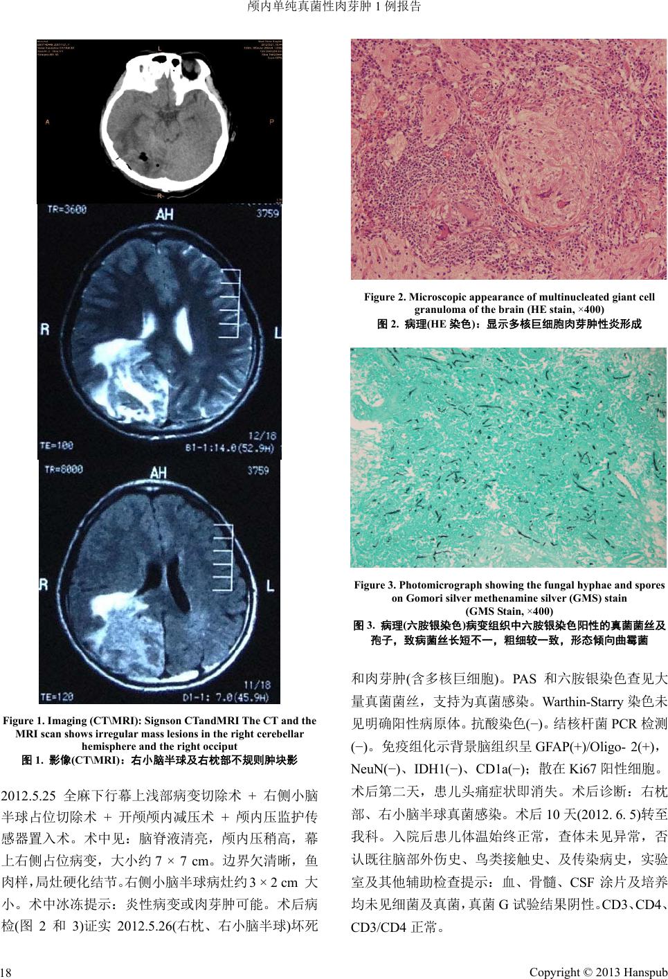 颅内单纯真菌性肉芽肿 1例报告 Figure 1. Imaging (CT\MRI): Signson CTandMRI The CT and the MRI scan shows irregular mass lesions in the right cerebellar hemisphere and the right occiput 图1. 影像(CT\MRI):右小脑半球及右枕部不规则肿块影 2012.5.25 全麻下行幕上浅部病变切除术 + 右侧小脑 半球占位切除术 + 开颅颅内减压术 + 颅内压监护传 感器置入术。术中见:脑脊液清亮,颅内压稍高,幕 上右侧占位病变,大小约 7 × 7 cm。边界欠清晰,鱼 肉样,局灶硬化结节。右侧小脑半球病灶约 3 × 2 cm 大 小。术中冰冻提示:炎性病变或肉芽肿可能。术后病 检(图2和3)证实 2012.5.26(右枕、右小脑半球)坏死 Figure 2. Microscopic appearance of multinucleated giant cell granuloma of the brain (HE stain, ×400) 图2. 病理(HE 染色):显示多核巨细胞肉芽肿性炎形成 Figure 3. Photomicrograph showing the fungal hyphae and spores on Gomori silver methenamine silver (GMS) stain (GMS Stain, ×400) 图3. 病理(六胺银染色)病变组织中六胺银染色阳性的真菌菌丝及 孢子,致病菌丝长短不一,粗细较一致,形态倾向曲霉菌 和肉芽肿(含多核巨细胞)。PAS 和六胺银染色查见大 量真菌菌丝,支持为真菌感染。Warthin-Starry 染色未 见明确阳性病原体。抗酸染色(−)。结核杆菌 PCR 检测 (−)。免疫组化示背景脑组织呈GFAP(+)/Oligo- 2(+), NeuN(−)、IDH1(−)、CD1a(−);散在Ki67 阳性细胞。 术后第二天,患儿头痛症状即消失。术后诊断:右枕 部、右小脑半球真菌感染。术后 10 天(2012. 6. 5)转至 我科。入院后患儿体温始终正常,查体未见异常,否 认既往脑部外伤史、鸟类接触史、及传染病史,实验 室及其他辅助检查提示:血、骨髓、CSF 涂片及培养 均未见细菌及真菌,真菌 G试验结果阴性。CD3、CD4、 CD3/CD4 正常。 Copyright © 2013 Hanspub 18  颅内单纯真菌性肉芽肿 1例报告 Copyright © 2013 Hanspub 19 2. 诊断 1) 颅内真菌性肉芽肿:曲霉菌感染?2) 幕上浅 部病变切除术 + 右侧小脑半 球占位切除术 后。术后 给予伏立康唑0.186~0.279 g Q12 h静滴 10 天,随后 更换为两性霉素B脂质体 4.5~140 mg Qd静滴 26 天, 停用两性霉素 B脂质体后给予伏立康唑 0.2~0.4 gbid 口服至术后2个月。至本文投送之日,患儿无临床症 状再次出现。 3. 讨论 真菌以不同方式和程度累及脑组织、脑膜和颅内 血管,其中颅内真菌性肉 芽肿(intracranial fungal granuloma, IFG)并不常见,但死亡率高(50%~ 63%)[1], 脑膜脑炎是最主要的死亡原因,感觉缺损、肉芽肿中 有脓性成分、严重脑水肿,均提示预后差。另外,实 质性肉芽肿常伴发血管炎和血管内血栓形成,一旦发 生,其死亡风险更高,考虑可能的原因是化学药物极 可能对难以逆转的血管内侵袭和继发性梗阻无效[2]。 但在儿童群体中的疾病结局极可能优于成人[3]。国内 IFG 文献多见于成人病例的个案报道,少见儿童报道。 IFG 最常见的症状是头痛、呕吐、凸眼、视力障碍及 视乳头水肿[1]。,临床症状及体征无特异性,CT、MRI 扫描往往无特征性表现,有文献提示CT 上的“开环 征”可能对诊断有一定的特异性[4],本患儿无CT 开 环征。病理染色可明确诊断真菌感染,但不能确认具 体病原,需要培养结果,但中枢神经系统感染真菌者, 脑脊液真菌培养阳性率往往并不高(31%)[5];病理确诊 的IFG 患者血/尿的涂片/培养的阳性率约 60%[3]。本 例患儿病理切片六胺银染色证实为真菌感染,结合临 床及病理表现特点病原菌可能为曲霉菌属(图2和3)。 曲霉菌是形成IFG 最常见的病原菌,IFG 感染途径主 要有:1) 直接感染:如开放性外伤感染;2) 血行播 散;3) 邻近感染:如鼻窦炎;4) 姻缘性。真菌感染 的易感因素包括营养状况差、滥用药物(抗生素、镇痛 药、抗结核、激素、免疫抑制剂)、高龄、患恶性肿瘤、 免疫功能缺陷和寄生虫感染等。没有发现可能的感染 途径和易感因素是本例患儿的特征之一。仅为限局性 肉芽肿并位于枕部、右小脑半球是本例患儿特征之 二,因IFG最常见部位为额叶、颞叶,其他部位为顶 叶、颞顶叶、前颅窝、中颅窝、鞍区和鞍旁区、后颅 窝[1]。本个案血、骨髓、CSF 涂片及培养均未见细菌 及真菌,真菌 G试验结果也为阴性。原因可能为:1) 该患儿除头痛、呕吐外,一般情况良好,除局灶占位 性改变外,并无大量真菌进入血液及其他体液中,故 出现上述病理与实验室检查结果的背离;2) 任何实验 室检查均可能有假阴性结果的存在。 手术联合抗真菌治疗是目前首选治疗方案。外科 病灶切除疗效确切,可减轻颅内压迫和降低颅内压, 防止发生严重的CNS 后遗症,改善预后。术后抗真 菌治疗应当早期、足量,足够疗程。初始全身治疗首 选伏立康唑,康唑不能耐受或治疗无效的患者,可替 代选用伊曲康唑、泊沙康唑或 LFABs(包括两性霉素B 脂质体复合物)。本例患儿手术后次日临床症状即完全 消失,随访至稿件投送之时,患儿无临床症状反复, 据此我们考虑是否应视为单纯手术效果好。该病例也 再次证明目前主流观点的正确性,即虽然没有决定性 的证据证明根治性和激进的手术能够促进颅内真菌 性肉芽肿患者的治疗结局,但是每当手术可行,且不 会导致严重的并发症等恶性结局时,仍应采取恰当而 彻底的手术[6]。 参考文献 (References) [1] A. R. Dubey, V. Patwardhan, S. Sampth, et al. Intracranial fungal granuloma: Analysis of 40 patients and review of the literature. Surgical Neurology, 2005, 63(3): 254-260. [2] B. S. Sharma, V. K. Khosla, V. K. Kak, et al. Intracranial fungal granuloma. Surgical, 1997, 47(5): 489-497. [3] J. Dotis,E. Iosifidis, E. Roilides, et al. Central nervous system aspergillosis in children: A systematic review of reported cases. International Journal of Infectious Diseases, 2007, 11(5): 381- 393. [4] 陈永华, 何玉麟, 李五根, 夏国金. 颅内真菌感染的 CT和 MRI 表现[J]. 中国医药导报, 2010, 7(17): 66-68. [5] C. Sundaram, P. Umabala, et al. Pathology of fungal infections of the central nervous system: 17 years’ experience from South- ern India. Histopathology, 2006, 49(4): 396-405. [6] A. A. Siddiqui, A. A. Shah and S. H. Bashir. Craniocerebrala- spergillosis of sinonasal origin in immunocompetent patients: Clinical spectrum and outcome in 25 cases. Neurosurgery, 2004, 55(3): 602-613. |