设为首页 加入收藏 期刊导航 网站地图
  • 首页
  • 期刊
    • 数学与物理
    • 地球与环境
    • 信息通讯
    • 经济与管理
    • 生命科学
    • 工程技术
    • 医药卫生
    • 人文社科
    • 化学与材料
  • 会议
  • 合作
  • 新闻
  • 我们
  • 招聘
  • 千人智库
  • 我要投搞
  • 办刊

期刊菜单

  • ●领域
  • ●编委
  • ●投稿须知
  • ●最新文章
  • ●检索
  • ●投稿

文章导航

  • ●Abstract
  • ●Full-Text PDF
  • ●Full-Text HTML
  • ●Full-Text ePUB
  • ●Linked References
  • ●How to Cite this Article
International Journal of Ecology 世界生态学, 2013, 2, 38-49
http://dx.doi.org/10.12677/ije.2013.24007 Published Online November 2013 (http://www.hanspub.org/journal/ije.html)
Development of Ecotoxicogenomic Biomarkers on the
Freshwater Shrimp (Neocaridina denticulate) Following
Short-Term Exposure to Dipropyl Phthalate
Y i-Chie Tsai, Hung-Hung Sung*
Department of Microbiology, Soochow University, Taipei
Email: *hhsung@scu.edu.tw
Received: Aug. 5th, 2013; revised: Sep. 10th, 2013; accepted: Sep. 26th, 2013
Copyright © 2013 Yi-Chie Tsai, Hung-Hung Sung. This is an open access article distributed under the Creative Commons Attribution License, which
permits unrestricted use, distribution, and reproduction in any medium, provided the original work is properly cited.
Abstract: Phthalate esters (PAEs), which are widely used in industrial chemicals that serve as important additives to
impart flexibility to polyvinyl chloride resins and have become widely diffused in the environment, are considered to be
endocrine disrupting chemicals (EDCs). In order to assess the toxicity of PAE to aquatic crustacean, after shrimps
(Neocaridina denticulate) were short-term exposed to a sublethal concentration (50 mg/L) of dipropyl phthalate (DPrP),
the differential expression genes were isolated and identified using suppression subtractive hybridization (SSH) on the
samples prepared from the whole individual. There were 71 unique expressed sequence tags (ESTs) which were identi-
fied by homology with data-based sequences, including 23 ESTs corresponded to known genes and 48 ESTs with un-
known function. The known genes could be divided into nine classes on the basis of physiological function: genes re-
lated to ribosomal, metabolism, immune and structural molecules, concerning the translation-related molecules, in-
volved respiration and signaling, and responding to signaling and vision. By comparing the level of gene transcription
in DPrP-treated group vs. non-treated group using semi-quantitative RT-PCR, we found that six of twelve selected
genes were significantly up-regulated following exposure to DPrP at high concentration (50 mg/L), including two im-
mune-related genes, one metabolism-related gene and 3 unknown genes. There were eight genes which are significantly
responding to DPrP treatment at 1.0 mg/L, including five up-regulated genes (four known genes and one unknown gene)
and three down-regulated unknown genes. After exposure to 0.5 mg/L of DPrP, only four genes (three known genes and
one unknown gene) were affected and their mRNA levels increased significantly. These results suggest that N. denticu-
late may be harmed via the change of the globally physiological function following exposure in non-lethal
PAE-polluted aquatic environment; in addition, the ESTs derived from N. denticulate can be used for studying the
ecotoxicological effect of other pollutants in the aquatic environment.
Keywords: Dipropyl Phthalate (Dprp); Endocrine Disrupting Chemical (EDC); Neocari dina denti cul at e; Aquatic
Crustacean; Differential Gene Expression; Suppression Subtractive Hybridization (SSH)
以短暂暴露于邻苯二甲酸二丙酯(Dipropyl Phthalate)之淡水
多齿新米虾(Neocaridina denticulata)建立生态毒理基因组生物
标志物
蔡伊茜,宋宏红*
东吴大学微生物学系,台北
Email: *hhsung@scu.edu.tw
收稿日期:2013 年8月5日;修回日期:2013年9月10 日;录用日期:2013 年9月26日
*通迅作者。
Open Access
38
以短暂暴露于邻苯二甲酸二丙酯(Dipropyl Phthalate)之淡水多齿新米虾
(Neocaridina denticulata)建立生态毒理基因组生物标志物
Open Access 39
摘 要:邻苯二甲酸酯类化合物(phthalate esters; PAEs)是广泛用于塑料制品之塑化剂和造成环境污染甚为可观的
內分泌干擾物質。本研究以多齿新米虾(Neocar i di na denti culata)暴露于次致死剂量(50 mg/L)之一種PAE——邻苯
二甲酸二丙酯(dipropyl phthalate; DPrP)后1天,利用抑制性扣减杂交法建立一套基因组指标,探讨其全面基因的
表现变化。实验中共筛得 71个独特的表达序列标签(expressed sequence tages; ESTs),包括 23段ESTs对应为已
知功能基因及48 段ESTs 为未知功能。根据生理功能,已知功能基因分别与 9类相关,包括代谢、呼吸、防御、
核糖体、染色体、转译、传讯、视觉与结构。虾子暴露于高浓度DPrP (50 mg/L)时,3个免疫及代谢相关的基因
和3个未知功能基因,共 6个基因的 mRNA 表现量明显下降。暴露于非致死剂量1.0 mg/L 时,共 8个基因受到
影响,包括正调节的 4个已知功能和 1个未知功能基因,以及 3未知功能基因被负调控。虾子暴露于更低剂量
0.5 mg/L 后,仅 4个基因受到影响,包括3个已知功能和 1个未知功能基因之mRNA 表现量明显上升。综合这
些结果暗示,包括 DPrP 在內之非致死浓度 PAEs 污染的水域可能藉由影响多齿新米虾的生理功能而威胁其存活;
此外,起源自多齿新米虾的ESTs也许可用在水生环境中其他污染物之生态毒性效应的研究。
关键词:邻苯二甲酸二丙酯(DPrP);内分泌干扰物质;多齿新米虾;水生甲壳类;差异表现基因;
抑制性扣减杂交法
1. 引言
内分泌干扰物质(Endocrine disrupting chemicals;
EDCs)是人造化学物,常经由 食物链 进入生 物体内 形
成假性荷尔蒙,使细胞误以为是化学讯号,干扰生物
体正常荷尔蒙的合成、分泌、传送及作用,进而影响
生物体的代谢、行为、生殖及性别分化等生理作用[1]。
这些外生性物质(xenobiotic compounds)的化性多安
定,在环境中不易被分解,易造成生物累积、生物浓
缩及生物转移;对暴露于其中的水生动物往往不具急
毒性(acute toxicity),但会导致慢性中毒而造成组织改
变。邻苯二甲酸酯类化合物(Phthalate esters; PAEs)为
制造塑料制品需添加的塑化剂,其种繁多且经常于制
造过程、使用和丢弃时释出至环境中[2],也曾在许多
动植物或肉制品中检测出[3]。在台湾,PAEs 广泛的存
在于河水、底泥和土壤中[4],并发现其累积在鱼体内
[5]。
许多实验发现,PAEs 因水溶性(solubility)和降解
性低,且亲脂性(hydroph obicity) 和吸附性高,可以在
陆地和水域环境的食物链中发生生物累积[2],造成不
正常的性发育和降低生殖功能而干扰繁殖和族群大
小[6-9]。Autian 研究指出,当母体内累积过多浓度的
PAE 会造成男性胎儿雌性化现象[10]。动物毒性试验研
究发现,某些 PAEs 对鼠類和大部份哺乳动物具有肝
脏和肾脏毒害性[11];PAEs 对鱼类(例如虹鳟、大西洋
鲑鱼等)具强烈之生物浓缩作用,易累积至生物体内之
脂肪层,长期下来会造成水中生物遗传因子突变、生
物体畸形、生理异常等[12,13]。目前 PAEs 已被多数国
家归类是一种非常普遍存在且优先列管控制的污染
物和 EDC。
许多无脊椎动物毒性测试议定书常规性地使用
在监管毒性测试[14]。其中淡水甲壳类具有许多优点,
包括在许多水域生态系中它们是主要的无脊椎动物、
族群个数经常众多且在实验室容易饲养。因此淡水甲
壳类目前被推荐用在监测环境污染状况[15-17]。多齿新
米虾(Neocar i d ina denticul ata)简称米虾,广泛分布在台
湾水域中,具有许多利于作为监测环境污染的优点,
包括体长仅 2至3 cm、喜栖息于干净、能自然繁殖、
且发育过程缺少变态期(metamorphosis stage)。自2005
年起,台湾环境保护署宣布米虾为一种急毒性测定的
指标生物,以评估各种水体或工厂排放水的质量。然
而,目前仅有少数研究是利用米虾以决定农药(如氯化
铵,硫酸铜,氯化镉,氯化汞,硫酸锌)的毒性[18]和氯
丹(Chlordane)及灵丹(Lindane)两种 EDCs 对生长和生
殖的影响[19,20]。本研究室曾将米虾浸泡于邻苯二甲酸
二丙酯(Dipropyl phthalate;DPrP)后,侦测六种不同的
生殖及免疫活性和一种免疫相关基因表现的变化,发
现次致死剂量的 DPrP会提高米虾的易感性(suscep-
tibility)[21],推测无致死剂量的 PAEs 仍可能对水域生
以短暂暴露于邻苯二甲酸二丙酯(Dipropyl Phthalate)之淡水多齿新米虾
(Neocaridina denticulata)建立生态毒理基因组生物标志物
态系造成潜在危机。因此,评估环境中 PAEs 的污染
及其毒性是非常重要的。然而针对PAEs 对水生生物
全面性生理之影响效应的相关研究极少。
近年来,以基因组学(genomics)、转录组学(trans-
criptomics)、蛋白质组学(proteomics)及代谢组学(meta-
bolomics)等藉由大规模方式以观察生物体内整体变
化进行研究的概念逐渐成熟,因为此研究方式能够提
供独有的信息。差异性表现基因组的研究适合用于观
察生物体内所有基因表现量变化的情形,足以系统性
评估生物体所受到的影响。目前常使用的方法之一是
抑制性扣减杂交法(suppressive subtractive hybridiza-
tion; SSH),主要原理是将两组样本的 mRNA 反转录
成cDNA 后进行杂合,剩余未杂合的样本即为表现有
差异的基因,接着以 PCR 放大表现有差异的基因且抑
制非目标基因的放大,达到扣减的效果。此方法能筛
选出表现量多和极少的基因[22]。Sung 等以生化指标和
基因指标评估不同浓度 DPrP 处理米虾不同时间后的
生殖和免疫毒性,发现基因表现对DPrP 为短期反应
且灵敏度优于生化活性[21]。因此推测米虾的基因组变
化可能具有作为水域环境之评估指标的潜力。
本研究目的在米虾暴露于 DPrP 后,建立一套基
因组指标,探讨 DPrP 处理后米蝦全面基因的表现变
化。实验中,由 DPrP 处理和未处理的两组虾子,利
用抑制性扣减杂交法筛选并鉴定出差异表现的基因
片段,即表达序列标签(expressed sequence tags; ESTs);
接着,选出数个目标基因,利用RT-PCR 法确认DPrP
对米虾生理功能的影响效应,并评估源自米虾的 ESTs
未来作为PAEs 污染指标的可能性。
2. 材料方法
2.1. 实验用虾及驯养
饲养虾子的玻璃缸(30 公分 × 30公分 × 30公分)
底部铺设厚约 3公分的美细砂,注入养殖池水并装置
外挂式过滤器,以过滤打气(150 L/小时)。三日后,放
入水草,并外加定时照明设备,水草每日照光八小时。
一周后,将购自一般水族馆的健康多齿新米虾
(Neocaridina denticulata)放至置备完成的池水中,养殖
密度约为每平方公尺 150~200 尾,养殖期间每日喂食
干燥孑孓一次,驯养三天后才进行实验。
2.2. 邻苯二甲酸二丙酯(dipropyl phthalate)的
制备
本研究室过去使用八种 PAEs实验证明,邻苯二
甲酸二丙酯(DPrP)对淡水长脚大虾(Macrobrachium
ro senb e rgii )免疫力的影响最为明显[23];米虾连续暴露
于不同剂量 DPrP 的10 天期间,于次致死剂量 50 m/L
处理后 1天的米虾各项免疫及生殖相关活性反应改变
最明显[21]。因此本研究选择将米虾浸泡于50 mg/L 之
DPrP,再进行后续实验的探讨。配制药物时,秤取 2500
mg 的DPrP,溶于 25 mL 的丙酮;再进行十倍稀释,
使浓度成为 105 mg/L和104 mg/L,置于室温下备用。
实验前,以丙酮调整浓度为2 × 10³ mg/L 和103 mg/L
后,再以养殖水调整 DPrP 浓度为 50 mg/L、1 mg/L
及0.5 mg/L;接着以三种浓度的池水分别进行后续不
同的实验。
2.3. 米虾的浸泡处理
为了得到讯息传导不同时期的差异表现基因,进
行抑制性扣减杂交前,米虾先浸泡于 50 mg/L 的DPrP
中2小时后,再转养至不含 DPrP 养殖池水后 12小时,
进行组织RNA 的萃取。为决定不同浓度 DPrP 对米虾
生理功能的影响,米虾分为三组实验组,分别浸泡于
50 mg/L、1 mg/L及0.5 mg/L,对照组米虾则浸泡于
不含 DPrP 池水。浸泡一天后,萃取米虾组织RNA。
2.4. RNA萃取与纯化
由于米虾过小,一只虾子难以收集足够的组织以
进行实验,所以本实验制备RNA 样本时,取相同处
理后的 10 只米虾为一个样本。以液态氮硏磨虾子后,
磨成粉状的组织放入 1毫升Trizol (Invitrogen, USA)
中,室温下作用 5分钟;接着,加入 0.2 毫升氯仿
(chloroform),剧烈震荡20 秒,于室温下静置 10 分钟;
再于 4℃下,以转速12,000  g,离心 15 分钟。取最
上层溶液,加入 1毫升异丙醇(isopropanol),均匀混合;
于室温下静置10分钟,再以 4℃,离心 15 分钟。去
除上清液后,加入 1毫升 75%酒精,清洗沉淀物,再
于4℃下,以转速 7500  g离心 5分钟。倒掉上清液,
风干 10 分钟后,取20 μL之DEPC 处理过的水以溶
解沉淀物,并于 55℃水浴 10分钟。最后,以核酸计
Open Access
40
以短暂暴露于邻苯二甲酸二丙酯(Dipropyl Phthalate)之淡水多齿新米虾
(Neocaridina denticulata)建立生态毒理基因组生物标志物
算仪(GeneQuant II, Phamacia Biotech, UK)定量 RNA
浓度,当 OD260/280为1.8~2.0 时,利用 mRNA 纯化试
剂(Invitrog en)以纯化 mRNA。
2.5. 抑制性扣减杂交(suppression subtractive
hybridization; SSH)
由实验组和控制组虾子得到的mRNA 样本后,利
用Clontech PCR-SelectTM cDNA扣减试剂和根据实验
手册进行抑制性扣减杂交(SSH)实验 ,以建立一个扣
减的 cDNA 基因库(library)。主要步骤简述如下。首
先进行第一股和第二股 cDNA 的合成,得到双股
cDNA。接着,以 RsaI限制脢切割双股 cDNA 两端成
钝端(blunt end),即进行接合子的连结。取1 μL切割
后的 DPrP 处理之实验组 cDNA,稀释后分取 2 μL置
于两个微量试管中,并分别于第一管加入 2 μL的接合
子adaptor 1,第二管加入adaptor 2R。在两管分别加
入接合酶(T4 DNA Ligase)溶液并混合均匀后,在 16
℃隔夜作用。作用后样本加入20X EDTA/Glycogen 终
止反应,并于72℃加热 5分钟,使 ligase 失去活性。
实验组样本制备完成后,开始进行杂合反应
(Hybridization) ,过程中共需进行两次杂合反应。首先,
取接合不同接合子的实验组样本各1.5 μL,分别加入
等体积经 Rsa I切割后之对照组 cDNA 样本和1 μL之
杂合缓冲液后,以 98℃作用90 秒,接着于 68℃作用
8小时,即完成第一次杂合。接着,取切割后对照组
cDNA 样本以98℃作用 90 秒后,加入第一次杂合的
两种不同接合子样本,混合均匀和稍做离心后,以 68
℃隔夜作用。反应后样本加入 200 μL的稀释缓冲液
(Clontech),以 68℃作用 7分钟后,即得到扣减后的
cDNA。
建立扣减cDNA 基因库前,扣减后cDNA 需先进
行两次的核酸脢连锁反应的放大(PCR Amplification)。
第一次加入PCR 引子 1、Taq DNA polymerase、10 mM
dNTP mixes和50 mM MgCl2以进行放大。第一次放
大的 PCR产物再以巢式 RCR (nested PCR)引子 1和巢
式PCR 引子 2R,进行第二次PCR 放大。本实验以
β-actin 为参考基因(GenBank accession no. AY947402)
[24] ,作为扣减过程是否完全的依据。接着,将PCR
产物接合至pGEM-T TA 质体后,加入胜任细胞
Escherischia coli JM109,于 37℃,转速 225 rpm 下,
震荡培养1.5 小时后,取100 L菌液涂抹培养于含有
X-gal、IPTG 及氨苄青霉素(ampicillin; 50 g/mL)的LB
培养基,建立扣减 cDNA基因库(subtracted cDNA
library)。为了检视培养基上的菌落内含有的载体具有
外来核酸片段,本实验利用巢式PCR 引子 1和巢式
PCR 引子2R,经 PCR后,放大产物以 2%琼酯胶体
法做确认。
另外,藉由限制酶切割图谱分析法以鉴别不同载
体是否具有不同外来核酸片段。使用常见三种限制酶
EcoRI、BamHI 和HimdIII (Biolabs)分别切割质体做,
经1%琼酯胶体电泳即得到的限制酶图谱。图谱片段
大小相同者视为同一群。在同一群内随机挑选数个样
本,送至阳明基因组中心进行扣减后核酸片段的定
序,再以 DNAStar 软件将有部分重复的序列进行前后
顺序的排列及重组。重组后序列于NCBI 数据库分别
以BLASTN 和BLASTX进行核酸序列及胺基酸序列
的比对。比对时,E值小于或等于 10−25 者,所得相似
序列定义为可信结果。
2.6. 半定量 RT-PCR分析
为了再次确认扣减后基因片段与 DPrP 处理的相
关性,由比对结果中选出八个已知功能及四个未知功
能的 ESTs (表1)作为分析的目标基因。根据不同ESTs
序列,设计个别的专一性的引子(表2),以半定量反转
录PCR 法分析 DPrP 处理后虾子之不同基因的 mRNA
表现量变化。暴露于不同浓度 DPrP之米虾组织
mRNA 后,加入合成cDNA 引子,70℃作用2分钟后,
加入 dNTP 和RNase 抑制剂(Fermentas)于37℃作用5
分钟;加入反转录酵素(20 units/μL; Fermentas),于42
℃作用 1小时,即完成cDNA 合成,即可进行放大。
本实验以 β-actin 作为内在控制组。放大后产物进行
1%的DNA 琼酯胶体电泳并照相后,以软件Image
Quant 5.1 分析产物条带的吸光强度,以相对吸收单位
(relative absorbance units)表示。同一样本中不同目标
基因表现量的半定量数值以比值(a ratio)表示,即某目
标基因的吸收单位除以同一样本的 β-actin的吸收单
位。本研究结果的图表数据则以某基因在实验组的比
值除以在控制组的比值表示(即a ratio of control)。
Open Access 41
以短暂暴露于邻苯二甲酸二丙酯(Dipropyl Phthalate)之淡水多齿新米虾
(Neocaridina denticulata)建立生态毒理基因组生物标志物
Open Access
42
Table 1. Characteristics of functional EST identified in subtractive libraries from DPrP-treated shrimp
表1. DPrP处理虾子后得到的扣减基因库中已知功能的表达序列标签性质
EST ID Gene identified (Accession No.) Function
groups Species Length of
fragment (bp)E value
Re1 16S rRNA (AY708114.1) 4 Caridina pristis 匙指虾 1425 0
Re2 16S rRNA (DQ463703.2) 4 Chloroflexi bacterium 绿弯菌 312 6.00E-41
Re4 Trypsin (X86369.1) 2 Litopenaeus vannamei 南美白虾 766 2.00E-96
Re5 Trypsin (AY596945.1) 2 Neocaridina denticulata sinensis 玫瑰虾 350 3.00E-77
Re6 Chymotrypsin 1 (X66415.1) 2 L. vannamei 南美白虾 306 8.00E-33
Re9 Hemocyanin (AF431737.1) 3,4 Penaeus monodon 草虾 760 1.00E-63
Re12 Opsin (Rh) gene (DQ852595.1) 5 Neomysis am ericana 黑褐新糠美洲虾 326 2.00E-92
Re13 Sarcoplasmic calcium-binding protein (SCP1)
(DQ256199.1) 6 Procambarus clarkii 克氏原螯虾 645 1.00E-78
Re14 Calcified cuticle protein (DQ288151.1) 7 Callinectes sa pidus 蓝蟹 608 4.00E-27
Re17 Elongation factor 1-alpha (AY117542.1) 8 Litopenaeus stylirostris 西方蓝虾 794 2.00E-59
Re18 Mitochondrial COI gene for cytochrome oxidase
subunit I (AB300187.1) 4 N. denticulata sinensis 玫瑰虾 693 9.00E-73
Re19 Collagenolytic serine protease (AF461035.1) 7 Paralithodes camtschaticus 石蟹 364 2.00E-42
Re20 Cathepsin-L-like cysteine peptidase (AY332270.1)2 Metapenaeus ensis 刀额新对虾 466 7.00E-36
DPrP1 Chromosome 4 (NW001263854.1) 9 Dictyostelium discoideum 黏菌 846 8.00E-32
DPrP9 QM protein (EU004069.1) 3 Marsupenaeus japonicus 日本对虾 398 1.00E-114
DPrP10 Phosphoenolpyruvate carboxykinase
(AJ132380.1) 2 Nephrops norvegicus 挪威龙虾 259 3.00E-43
DPrP11 Mitochondrion (EF584003.1) 4 L. vannamei 南美白虾 612 1.00E-69
DPrP15 Myosin light chain (HM034314.1) 7 Penaeus monodon 草虾 507 5.00E-37
DPrP16 Mitochondrial cytochrome oxidase subunit 2
(COII) (AY842663.1) 4 Hemipyrellia ligurriens 瘦叶带绿蝇 270 5.00E-47
DPrP21 Preamylase 1 (X77318.1) 2 L. vannamei 南美白虾 251 8.00E-32
DPrP24 ribosomal protein 4 (AY829788.1) 1 Lonom i a o blique 天蚕蛾幼虫 254 4.00E-42
DPrP27 ribosomal protein L8 (DQ316258.1) 1 L. vannamei 南美白虾 236 7.00E-64
DPrP31 S2 ribosomal protein (AY292479.1) 1 Eleginus gracilis 红花鳕鱼 125 5.00E-30
The number in the third column indicates functional groups. 1: Ribosomal, 2: Metabolism, 3: Immune response, 4: Respiration, 5: Vision, 6: Signaling, 7: Structure, 8:
Translation, 9: Chromosome.
2.7. 统计分析
由于米虾非纯品系(outbred)动物,个体之间表现
型背景的差异性大,所以每一相对数值至少进行 5重
复,每一重复至少使用 5只虾子。以 one-way ANOVA
和Duncan进行不同浓度处理组之间的比较,分析后
有显着差异者(P < 0.05)。
3. 结果
3.1. 差异表现片段的扣减基因库(subtracted
cDNA library)的建立
为了得到 DPrP 处理前后具有差异异性表现之基
因,本研究由实验组及控制组的虾子,利用抑制性扣
减杂交法后,针对所有转型株,利用巢式 PCR 确认后,
筛选获得431 个转型株。进一步确认插入之基因片段
是否相同,以三种限制酶EcoRI、Bam HI 和HimdⅢ进
行切割,以分析各 cDNA 的限制图谱后,共挑选得 226
个转型株,即得到一个扣减后cDNA 基因库。
3.2. 基因片段的比对
基因库所有基因片段定经序后,以 DNAStar 软件
将不同片段序列上重复的部分进行前后顺序的排列
及重组,完成后得到共获得71 个独特的表达序列标
签(expressed sequence tages; ESTs)。接着,利用 NCBI
以短暂暴露于邻苯二甲酸二丙酯(Dipropyl Phthalate)之淡水多齿新米虾
(Neocaridina denticulata)建立生态毒理基因组生物标志物
Table 2. The specific primers of ESTs used in this study
表2. 使用在本研究之表达序列标签的专一性引子序列
EST ID Sequence for primers (5’3’) Size (bp)
Re9 L:CAGCCCAAATGTCCGTTACT
R:CCTTGAAGCTTGGCACATC 600
Re17 L:CGTGAGCACGTCTTGCTTT
R:TGAGCAGTGTGGCAGTCAA 701
Re18 L:GGCTCGTGTGTCAACATCC
R:CCCCCATTAGCAAGAGGAA 522
R20 L:CTTCAACCCAGCCAATGTG
R:GGTAGCAATGCCGCAGTTA 337
DPrP9 L:GCC TTGTTACGGTCGTGAGT
R:GGGAAGAAAGAAGGCTGACG 330
DPrP10 L:CTCATGTCCCAGAAACCAA
R:CCCAGGTAAAGAAGGAGTCA 248
DPrP11 L:CACACCTCGCGGGTAGTAGT
R:ACCAAGCTGCTGCTTCAAA 510
DPrP15 L:CCTCCAGGTCTTCTGGTGAG
R:GAAGCGCTTGAGTGTCACAG 375
uDPrP4 L:ATGTCGAGTGCCAGCTCAG
R:GTGTTGGCAGTGACGACTCA 315
uDPrP12 L:CTTCAGCAACTGCAAGTCCA
R:GCATGACTGTGAGGGCTTTT 356
uDPrP13 L:CACTAGGCGAGTTAAGGATG
R:GTTTCCTTTATGGTGCGAGT 254
uDPrP14 L:GCCGATGTCACGGATAAGTC
R:GGAGTCTGT GCCCAAGTCAT 304
EST, expressed sequence tag; uDPrP denotes ESTs had not be identified in the
public databases and are regarded as function unknown genes.
的BLASTN 和BLASTX分别进行核酸序列及胺基酸
序列的比对,将其中核酸序列比对所得之E值低于(或
等于)10−25 且比对到的物种为无脊椎动物之基因片
段,定义为已知生理功能;比对后得到 23 段ESTs 对
应为已知功能基因(表1)及48段ESTs为未知功能。
已知功能ESTs 大致可分与九类生理功能相关,包括:
核糖体组成的核糖体蛋白(ribosomal proteins;DPrP24,
DPrP27 及DPrP31);代谢相关基因的胰蛋白酶(trypsin;
Re4 及Re5)、胰凝乳蛋白酶(chymotrypsin; Re6)、淀 粉
脢原(preamylase; DPrP21)、溶脢体组织蛋白脢
(cathepsin-L-like cysteine peptidase; Re20)和磷酸烯醇
式丙酮酸羧激酶(Phosphoenolpyruvate carboxykinase;
DPrP10);防御相关的血青素(hemocyanin; Re9)、QM
蛋白(QM protein; DPrP9);呼吸相关的粒线体基因
(DPrP11)、粒线体16S rRNA (Re1及Re2)、血青素
(Re9)、细胞氧化酵素COI (cytochrome oxidase subunit
I; Re18)及COII (cytochrome oxidase subunit II;
DPrP16);视觉相关的视紫蛋白(opsin; Re12);讯号 传
递相关的携钙蛋白(sarcoplasmic calcium-binding pro-
tein; Re13);细胞和组织结构相关 的胶 原溶 解丝 氨酸
蛋白酶(Collagenolytic serine protease; Re19)和肌球蛋
白轻链(Myosin light chain; DPrP15);转译相关的延长
因子(elongation factor 1-alpha; Re17)与染色体相关基
因(Chromosome 4; DPrP1)等(表1)。
3.3. 不同浓度 DPrP 对目标基因的影响
确认上述得到的 ESTs 是否因 DPrP 处理而表现确
有差异,实验中选取不同已知功能的ESTs 和四个未
知功能 ESTs,以 RT-PCR半定量法,对暴露于不同浓
度DPrP 的米虾,进行这些目标基因之 mRNA 合成量
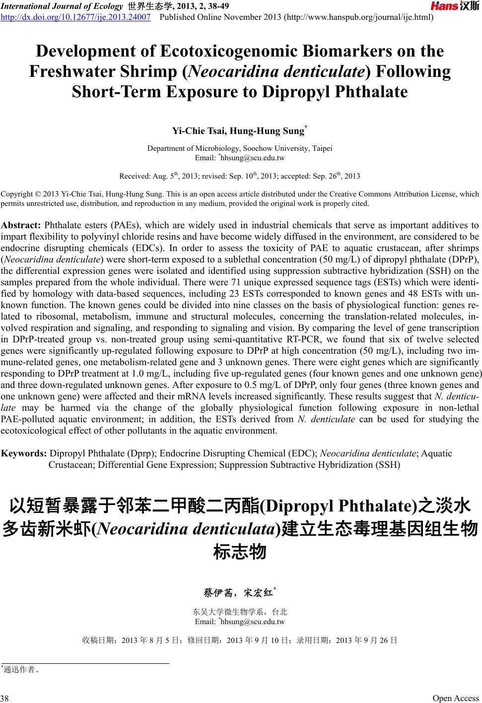
的分析。DPrP 影响免疫相关基因的结果如图 1所示,
浓度为 0.5~1.0 mg/L会明显增进血青素基因(hc)
mRNA 量,对MQ 基因(mq)则无明显影响;DPrP浓
度提高至50 mg/L 明显抑制两个基因的转录。对于代
谢相关基因,DPrP 于0.5 和1.0 mg/L 时会促进溶脢体
组织蛋白脢基因catl 的表现,浓度为50 mg/L 的DPrP
对此基因无影响;然而,基因磷酸烯醇式丙酮酸羧激
Re9
Hemocyanin
Ratio of control
0.0
0.5
1.0
1.5
2.0
2.5
3.0
*
*
*
DPrP7
QM protein
Ratio of control
0.0
0.5
1.0
1.5
2.0
2.5
3.0
*
0.51.0 50.0
Concentration (mg/L)
Figure 1. Messenger RNA expression of immunity-related genes of
Neocaridina denticulate treated with various concentrations of
dipropyl phthalate (DPrP) for one day
图1. 米虾曝露于不同浓度之邻苯二甲酸二丙酯一天后免疫相关基
因的讯息 RNA 表现量
Open Access 43
以短暂暴露于邻苯二甲酸二丙酯(Dipropyl Phthalate)之淡水多齿新米虾
(Neocaridina denticulata)建立生态毒理基因组生物标志物
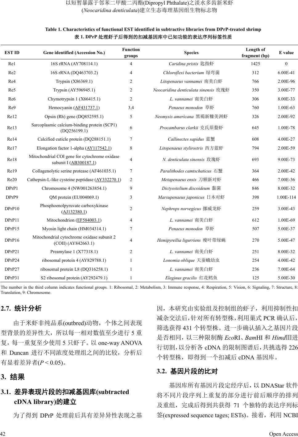
酶pepck 仅在米虾暴露于 50 mg/L时受到影响,mRN A
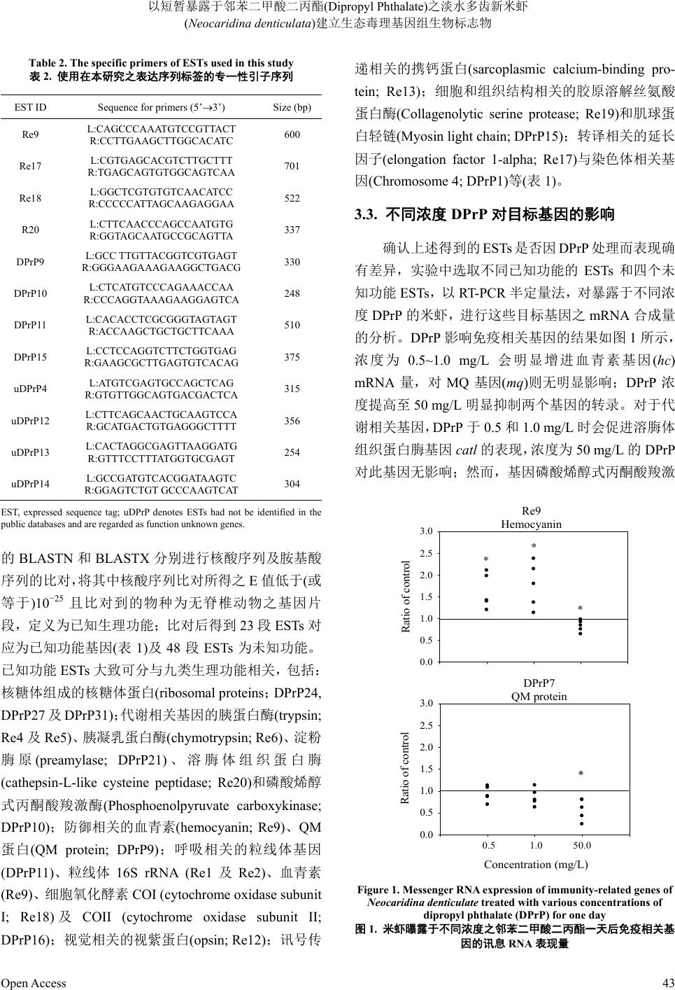
量明显下降(图2)。DPrP 除了在 1 mg/L 会增加粒线体
基因 mit 的表现外,其他浓度对两个呼吸相关基因mit
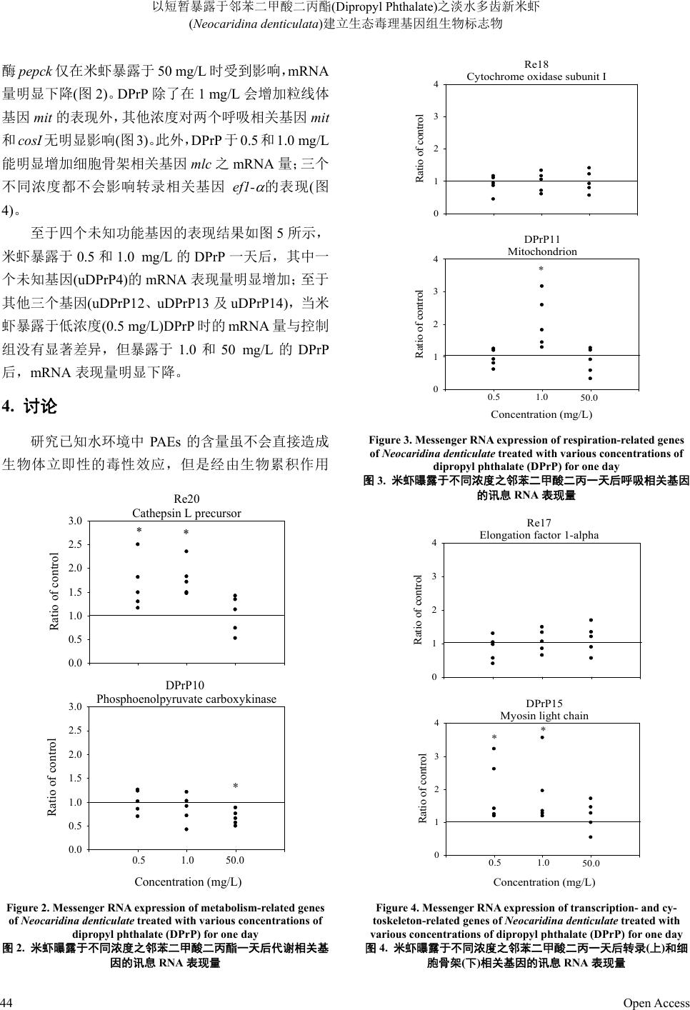
和cosI无明显影响(图3)。此外,DPrP 于0.5 和1.0 mg/L
能明显增加细胞骨架相关基因mlc 之mRNA 量;三个
不同浓度都不会影响转录相关基因 ef1-

的表现(图
4)。
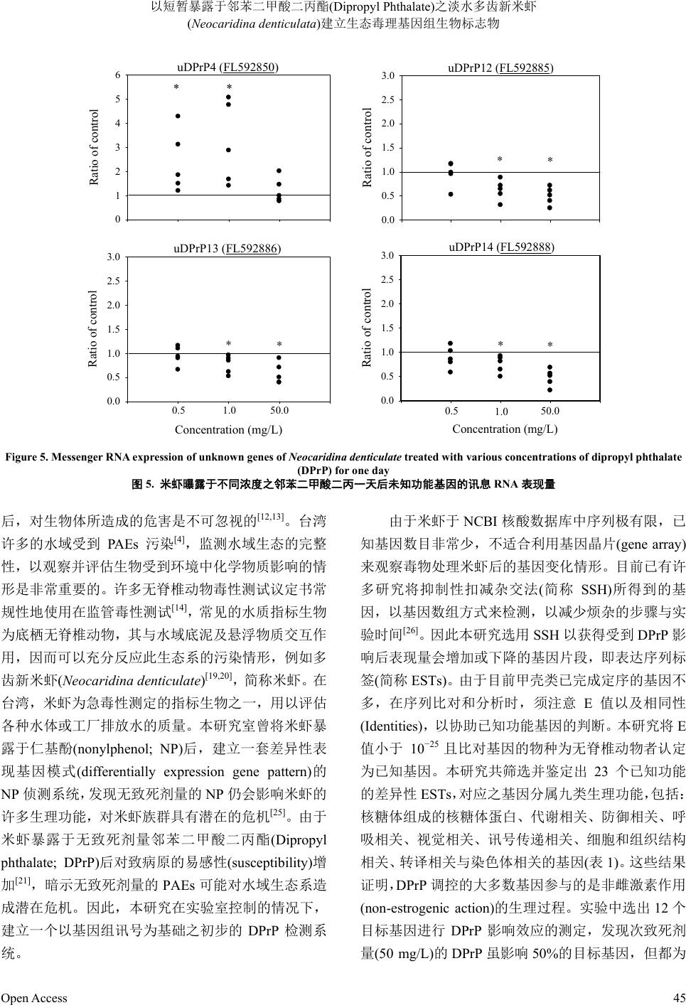
至于四个未知功能基因的表现结果如图 5所示,
米虾暴露于 0.5 和1.0 mg/L的DPrP 一天后,其中一
个未知基因(uDPrP4)的mRNA 表现量明显增加;至于
其他三个基因(uDPrP12、uDPrP13 及uDPrP14),当 米
虾暴露于低浓度(0.5 mg/L)DPrP时的 mRNA 量与控制
组没有显著差异,但暴露于1.0和50 mg/L的DPrP
后,mRNA 表现量明显下降。
4. 讨论
研究已知水环境中 PAEs的含量虽不会直接造成
生物体立即性的毒性效应,但是经由生物累积作用
DPrP10
Phosphoenolpyruvate carboxykinase
Concentration (mg/L)
Ratio of control
0.0
0.5
1.0
1.5
2.0
2.5
3.0
Re20
Cathepsin L precursor
Ratio of control
0.0
0.5
1.0
1.5
2.0
2.5
3.0
**
*
0.51.0 50.0
Figure 2. Messenger RNA expression of metabolism-related genes
of Neocaridina denticulate tr eated with various concentrations of
dipropyl phthalate (DPrP) for one day
图2. 米虾曝露于不同浓度之邻苯二甲酸二丙酯一天后代谢相关基
因的讯息 RNA 表现量
Re18
Cytochrome oxidase subunit I
Ratio of control
0
1
2
3
4
DPrP11
Mitochondrion
Ratio of control
0
1
2
3
4
*
Concentration (mg/L)
0.51.050.0
Figure 3. Messenger RNA expression of respiration-related genes
of Neocaridina denticulate treated with various concentrations of
dipropyl phthalate (DPrP) for one day
图3. 米虾曝露于不同浓度之邻苯二甲酸二丙一天后呼吸相关基因
的讯息 RNA 表现量
Re17
Elongation factor 1-alpha
Ratio of control
0
1
2
3
4
DPrP15
Myosin light chain
0
1
2
3
4
*
Concentration (mg/L)
*
0.51.0 50.0
Ratio of control
Figure 4. Messenger RNA expression of transcription- and cy-
toskeleton-related genes of Neocaridina denticulate treated with
various concentrations of dipropyl phthalate (DPrP) for one day
图4. 米虾曝露于不同浓度之邻苯二甲酸二丙一天后转录(上)和细
胞骨架(下)相关基因的讯息 RNA表现量
Open Access
44
以短暂暴露于邻苯二甲酸二丙酯(Dipropyl Phthalate)之淡水多齿新米虾
(Neocaridina denticulata)建立生态毒理基因组生物标志物
Open Access 45
uDPrP4 (FL592850)
Ratio of control
0
1
2
3
4
5
6
**
uDPrP13 (FL592886)
Concentration (mg/L)
Ratio of control
0.0
0.5
1.0
1.5
2.0
2.5
3.0
0.5 1.0 50.0
*
*
uDPrP12 (FL592885)
Ratio of control
0.0
0.5
1.0
1.5
2.0
2.5
3.0
*
*
uDPrP14 (FL592888)
Concentration (mg/L)
Ratio of control
0.0
0.5
1.0
1.5
2.0
2.5
3.0
**
0.5 1.0 50.0
Figure 5. Messenger RNA expression of unknown genes of Neocaridina denticulate treated with various concentrations of dipropyl phthalate
(DPrP) for one day
图5. 米虾曝露于不同浓度之邻苯二甲酸二丙一天后未知功能基因的讯息RNA表现量
后,对生物体所造成的危害是不可忽视的[12,13]。台湾
许多的水域受到 PAEs 污染[4],监测水域生态的完整
性,以观察并评估生物受到环境中化学物质影响的情
形是非常重要的。许多无脊椎动物毒性测试议定书常
规性地使用在监管毒性测试[14],常见的水质指标生物
为底栖无脊椎动物,其与水域底泥及悬浮物质交互作
用,因而可以充分反应此生态系的污染情形,例如多
齿新米虾(Neocaridina denticulate)[19,20],简 称 米 虾 。在
台湾,米虾为急毒性测定的指标生物之一,用以评估
各种水体或工厂排放水的质量。本研究室曾将米虾暴
露于仁基酚(nonylphenol; NP)后,建立一套差异性表
现基因模式(differentially expression gene pattern)的
NP 侦测系统,发现无致死剂量的 NP 仍会影响米虾的
许多生理功能,对米虾族群具有潜在的危机[25]。由于
米虾暴露于无致死剂量邻苯二甲酸二丙酯(Dipropyl
phthalate; DPrP)后对致病原的易感性(susceptibility)增
加[21],暗示无致死剂量的PAEs可能对水域生态系造
成潜在危机。因此,本研究在实验室控制的情况下,
建立一个以基因组讯号为基础之初步的 DPrP 检测系
统。
由于米虾于 NCBI 核酸数据库中序列极有限,已
知基因数目非常少,不适合利用基因晶片(gene array)
来观察毒物处理米虾后的基因变化情形。目前已有许
多研究将抑制性扣减杂交法(简称 SSH)所得到的基
因,以基因数组方式来检测,以减少烦杂的步骤与实
验时间[26]。因此本研究选用SSH 以获得受到 DPrP 影
响后表现量会增加或下降的基因片段,即表达序列标
签(简称 ESTs)。由于目前甲壳类已完成定序的基因不
多,在序列比对和分析时,须注意E值以及相同性
(Identities),以协助已知功能基因的判断。本研究将E
值小于 10−25 且比对基因的物种为无脊椎动物者认定
为已知基因。本研究共筛选并鉴定出 23个已知功能
的差异性ESTs,对应之基因分属九类生理功能,包括:
核糖体组成的核糖体蛋白、代谢相关、防御相关、呼
吸相关、视觉相关、讯号传递相关、细胞和组织结构
相关、转译相关与染色体相关的基因(表1)。这 些结 果
证明,DPrP 调控的大多数基因参与的是非雌激素作用
(non-estrogenic action)的生理过程。实验中选出 12 个
目标基因进行 DPrP 影响效应的测定,发现次致死剂
量(50 mg/L)的DPrP 虽影响50%的目标基因,但都为
以短暂暴露于邻苯二甲酸二丙酯(Dipropyl Phthalate)之淡水多齿新米虾
(Neocaridina denticulata)建立生态毒理基因组生物标志物
向下调节效应(down-regulation effect),显示对米虾生
理作用产生相当的压迫。当暴露剂量降低至无致死剂
量的 1 mg/L时,67%目标基因受到 DPrP 的上调或下
调,显示影响米虾的生理层面广;若暴露于更低浓度
0.5 mg/L 时,仅33%目标基因受到影响,且均为上调
效应(up-regulation effect)。这些无致死剂量 DPrP 对米
虾基因的影响结果无法判断是否对米虾的生理功能
具有压迫性,需进一步探讨。此外,米虾暴露在本实
验使用的三种次致死或无致死浓度之DPrP 后,在 12
个选定的目标基因中,讯号传递相关的 ef1-

和呼吸
相关的 cosI两个基因完全不受到影响;但其他主要参
与免疫(hc、qm和catl)和代谢(catl 和pepck)相关的基
因则对不同浓度DPrP 有不同反应(表3)。已知米虾暴
露于无致死剂量邻 DPrP 后的易感性(susceptibility)增
加[21],结合本研究结果推测,水体出现无致死剂量的
PAEs 可能主要经由防御或代谢路径影响米虾族群的
存活。
对大多数的甲壳类而言,污染物造成的环境压力
似乎是引起免疫力下降及患病率增加的重要因子之
一[27,28]。先前的研究显示,PAEs会降低米虾和淡水长
脚大虾的免疫活性和增加易感性[21,23];本研究证明 hc
及qm的表现确实会受 DPrP 调节,尤其高浓度 DPrP
会降低两个基因的表现。在节肢动物,血清素
(hemocyanin; Hc)是在肝胰脏合成[29,30],且为六聚合体
(hexamer)的氧气载体(oxygen carrier)。除了携带氧气
的功能外,许多研究也证明 Hc具有防御功能,例如
蜘蛛(Eurypelma californicum)[31-34]和鲎(Tachypleus-
tridentatus)[35]的Hc 经特定蛋白质水解后具有酚氧化
酵素(phenoloxidase)活性,且甲壳类的 Hc 经蛋白酵素
分解后,产生具抗微生物活性的抗微生物胜肽
(antimicrobial peptides)[36,37]。另一个免疫相关基因qm
的产物 QM 蛋白质,其在哺乳类和酵母菌细胞是一种
核糖体蛋白质[38,39];更多证据发现,QM 类蛋白
(QM-like protein)在细胞生长和分化等时期具有核糖
体蛋白质以外的功能[40,41]。Xu 等人以 SSH 方法分析
得知,白点病毒感染之日本对虾(Penaeus japonicas)
体内的 QM表现大量增加,且能够与Hc和肌球蛋白
(myosin)形成复合物而参与原酚氧化酵素系统(简称
proPO系统)和调节 Hc的酚氧化酵素活性,显示 QM
在节肢动物免疫系统具有特殊功能[42]。本研究室先前
Table 3.The summary of the effect of dipropyl phthalate (DPrP) on
the EST expression of Neocaridina denticulata
表3. 不同浓度邻苯二甲酸二丙酯基对米虾表达序列标签表现的影
响效应之摘要
DPrP (mg/L)
EST ID Target genes
0.5 1.0 50
Immunity
Re9 Hemocyanin (hc) E E D
DPrP9 QM protein (qm) N N D
Metabolism
R20 Cathepsin-L-like cysteine
peptidase (catl) E E N
DPrP10 Phosphoenolpyruvate
carboxykinase (pepck) N N D
Translation
Re17 Elongation factor 1-alpha (ef1-

) N N N
Respiration
Re18 Mitochondrial cytochrome
oxidase subunit I (cos I) N N N
DPrP11 Mitochondrion (mit) N E N
Cytoskeleton
DPrP15 Myosin light chain (mlc) E E N
Unknown function
uDPrP4 E E N
uDPrP12 N D D
uDPrP13 N D D
uDPrP14 N D D
Abbreviations are as follows: E = an enhancement effect of NP on gene expres-
sion; D = a reduction effect of NP gene expression; and N = no difference of gene
expression in DPrP-treated individuals from that in corresponding control.
研究指出,DPrP会影响米虾的超氧岐化酵素(super-
oxide dismutase; SOD)和原酚氧化酵素(prophenoloxi-
dase)等免疫酵素活性[21]。本研究结果推测,DPrP 也
可能透过影响hc和qm 的表现而而参与 proPO 系统以
调控米虾的免疫力。
细胞自溶酵素(cathepsin L; CatL)属于硫醇型蛋白
质分解酵素(cysteine protease)家族,多存在于胞内溶
体(lysosome)中,广泛的分布于正常组织,在发育过
程扮演重要角色,主要参与分化、原肠型成、脱皮及
卵黄利用等作用[43-49]。在无脊椎动物,硫醇型蛋白质
分解酵素与细胞内蛋白质的分解有关[50]。在甲壳类,
Open Access
46
以短暂暴露于邻苯二甲酸二丙酯(Dipropyl Phthalate)之淡水多齿新米虾
(Neocaridina denticulata)建立生态毒理基因组生物标志物
CatL 主要存在于肝胰脏,参与食物的分解,例如南美
白对虾(Litopenaeus vannamei)[51]和刀额新对虾 (Meta-
penaeus ensi s)[52]。近年研究证明,CatL 参与无脊椎动
物的先天性免疫力,包括南美白对虾[53]、中国对虾
(Fenneropenaeus chinensis)[54]、中华绒螯蟹(Eriocheir
sinensis)[55]和合浦珠母贝(Pinctada fucata)[56]等。根据
这些研究推测,DPrP 对米虾的防御反应或代谢活性具
有多重调节效应(multi-modulating effect)。相同的现象
也发生在NP 对米虾的影响,但是调节效应相反[25]。
磷酸烯醇式丙酮酸羧激酶(Phosphoenolpyruvate
carboxykinase)是醣质新生(gluconeogenesis)过程的重
要酵素,负责将草醋酸(oxaloacetate)转换成磷酸烯醇
丙酮酸(phosphoenolpyruv ate)和二氧化碳,以供后续转
换为葡萄糖。在某些特定的组织或细胞中相当重要,
例如大脑、肾脏髓质等皆以葡萄糖作为唯一能量来
源,当葡萄糖量不足时,就必须以醣质新生合成葡萄
糖来维持正常生理。本研究发现此酵素基因 pepck的
表现仅在高浓度时会受到抑制,推测次致死剂量可能
影响葡萄糖的恒定而对米虾造成一定程度的压迫。
综合以上暴露于 DPrP 后差异性表现基因的表现
結果证明,DPrP 主要经由多重生理功能的改变对米虾
产生外生毒力(ectotoxigenicity),推测无致死剂量 PAE
的污染仍可能危害到米虾族群的生存。由于台湾的环
境保护署使用米虾以监测水体的急毒性,结合本研究
的结果建议,米虾确实可作为水域指针系统(aquatic
indicator system)以评估水体污染对水生环境或生物的
风险,且发展自米虾的差异性表现ESTs未来应具有
作为基因组生物标志(genomic biomarkers)的潜力。由
于本研究获得的有效 ESTs 种类有限,因此未来仍须
搜集和鉴定更多污染相关的专一性基因,并整合更多
针对不同毒物的差异反应信息,以建立一套更具灵敏
度及可信度的监测模式组。
5. 致谢
本研究感谢台湾的国家科学委员会之计划经费
支持(NSC96-2745-B-031-003-URD)。
参考文献 (References)
[1] Holmes, P., Harrison, P., Bergman, A., Brouwer, I., Brouwer, B.,
Keiding, N., Randall, G., Sharpe, R., Skakkebaek, N., Ashby, J.,
Barlow, S., Dickerson, R., Humfrey, C. and Smith, L.M. (1997)
European workshop on the impact of endocrine disrupters on
human and wildlife. Proceedings of a Workshop, Weybridge, UK,
Report NO. EUR 17549.
[2] Staples, C.A., Peterson, D.R., Parkerton, T.F. and Adams, W.J.
(1997) The environmental fate of phthalate esters: A literature
review. Chemosphere, 35, 667-749.
[3] Yin, M.C. and Su, K.H. (1996) Investigation on risk of phthalate
ester in drinking water and marketed foods. Journal of Food and
Drug A n al ys is , 4, 313-318.
[4] Liu, C., Wang, S.K. and Lu, Y.B. (2000) Chemical characteriza-
tion of Tamshui River sediment in northern Taiwan. Journal of
Food and Drug Analysis, 76, 205-218.
[5] Chang, B.V., Liao, C.S., Huang, B.B. and Lee, C.C. (2004)
Occurrence of phthalate esters in Taiwan. Proceeding of the
Third Conference on Environmental Hormones and POPs, Tai-
pei, 12 November 2004, 63-66.
[6] Eckardt, R.E. (1973) Recent developments in industrial car-
cinogens. Journal of Occupational Medicine, 15, 904-907.
[7] Tavares, I.A. and Vine, N.D. (1985) Phthalic acid esters inhibit
arachidonate metabolism by rat peritoneal leucocytes. Journal of
Pharmacy and Pharmacology, 37, 67-68.
[8] Carozzi, S., Nasini, M.G., Schelotto, C., Caviglia, P.M., Santoni,
O. and Pietrucci, A. (1993) A biocompatibility study on perito-
neal dialysis solution bags for CAPD. Advances in Peritoneal
Dialysis, 9, 138-142.
[9] Jha, A.M., Singh, A.C. and Bharti, M. (1998) Germ cell muta-
genicity of phthalic acid in mice. Mutation Research, 422, 207-
212.
[10] Autian, J. (1973) Toxicity and health threats of phthalate esters:
Review of the literature. Environmental Health Perspectives, 4,
3-26.
[11] Thomas, J. and William, L. (1975) Biodegradation of di-n-butyl
phthalate and di-2-ethylhexyl phthalate in freshwater hydrosoil.
Journal of Fisheries Science and Technology, 32, 333-339.
[12] Capacchietti, M., Sabbieti, M.G., Materazzi, S., Materazzi, G.,
Menghi, G. and Marchetti, L. (2007) Phthalate esters immu-
nolocalized in the gastrointestinal tract of shi drum, Umbrina
cirrosa (L.) and rainbow trout, Oncorhynchus mykiss (W.). His-
tology and Histopathology, 22, 15-21.
[13] Norman, A., Borjeson, H., David, F., Tienpont, B. and Norrgren,
L. (2007) Studies of uptake, elimination, and late effects in At-
lantic salmon (Salmo salar) dietary exposed ot di-2-ethylhexyl
phthalate (DEHP) during early life. Archives of Environmental
Contamination and Toxicology, 52, 235-242.
[14] Schweer, L.G. (2002) Draft detailed review paper on mysid life
cycle toxicity test. US Environmental Protection Agency, Wash-
ington DC.
[15] Shigehisa, H. and Shiraishi, H. (1998) Biomonitoring with
shrimp to detect seasonal change in river water toxicity. Envi-
ronmental Toxicology and Chemistry, 17, 687-694.
[16] Fossi, M.C., Marsili, L., Neri, G., Casini, S., Bearzi, G., Politi, F.,
Zanardelli, M. and Panigada, S. (2000) Skin biopsy of mediter-
ranean cetaceans for the investigation of interspecies suscepti-
bility to xenobiotic contaminants. Marine Environmental Re-
search, 50, 517-521.
[17] Kirkpatrick, A.J., Gerhardt, A., Dick, J.T.A., McKenna, M. and
Berges, J.A. (2006) Use of the multispecies freshwater biomonitor
to assess behavioral changes of Corophium volutator (Pallas,
1766) (Crustacea, Amphipoda) in response to toxicant exposure
in sediment. Ecotoxicology and Environmental Safety, 64, 298-
303.
[18] Kitamura, H. (1990) Relation between the toxicity of some
toxicants to the aquatic animals (Tanichthys albonubes and
Neocaridina denticulata) and the hardness of the test. Bulletin of
the Faculty of Fisheries Nagasaki University, 67, 13-19.
[19] Huang, D.J. and Chen, H.C. (2004) Effects of chlordane and
lindane on testosterone and vitellogenin levels in green neon
Open Access 47
以短暂暴露于邻苯二甲酸二丙酯(Dipropyl Phthalate)之淡水多齿新米虾
(Neocaridina denticulata)建立生态毒理基因组生物标志物
shrimp (Neocaridina denticulate). International Journal of Toxi-
cology, 23, 91-95.
[20] Huang, D.J., Chen, H.C., Wu, J.P. and Wang, S.Y. (2006) Re-
production obstacles for the female green neon shrimp (Neo-
caridina denticulate) after exposure to chlordane and lindane.
Chemosphere, 64, 11-16.
[21] Sung, H.H., Lin, Y.H. and Hsiao, C.Y. (2011) Potential toxicity
of dipropyl phthalate to physiological functions of the green
neon shrimp (Neocaridina denticulate). Fish & Shellfish Immu-
nology, 31, 511-515.
[22] Larkin, P., Knoebl, I. and Denslow, N.D. (2003) Differential
gene expression analysis in fish exposed to endocrine disrupting
compounds. Comparative Biochemistry and Physiology Part B:
Biochemistry and Molecular Biology, 136, 149-161.
[23] Sung, H.H., Kao, W.Y. and Su, Y.J. (2003) Effects and toxicity
of phthalate esters to hemocytes of giant frechwater prawn,
Macrobranchium rosenbergii. A qua tic Toxicology, 64, 25-37.
[24] Lu, K.Y., Huang, Y.T., Lee, H.H. and Sung, H.H. (2006) Cloning
the prophenoloxidase cDNA and monitoring the expression of
proPO mRNA in prawns (Macrobrachium rosenbergii) stimu-
lated in vivo by CpG oligodeoxynucleotides. Fish & Shellfish
Immunology, 20, 274-284.
[25] Liu, C.L. and Sung, H.H. (2011) Genes are differentially ex-
pressed at transcriptional level of Neocaridina denticulata fol-
lowing short-term exposure to nonylphenol. Bulletin of Envi-
ronmental Contamination and Toxicology, 87, 220-225.
[26] Feng, L., Miao, W. and Wu, Y. (2007) Differentially expressed
genes of Tetrahymena thermophila in response to tributyltin
(TBT) identified by suppression subtractive hybridization and
real time quantitative PCR. Aqua ti c Toxicology , 81, 99-105.
[27] Victor, B., Narayanan, M. and JonesNelson, D. (1990) Gill pa-
thology and hemocyte response in mercury exposed Macro-
brachium idae (Heller). Journal of Environmental Biology, 11,
61-65.
[28] Sindermann, C.J. (1979) Pollution-associated diseases and ab-
normalities of fish and shellfish: A review. Fishery Bulletin, 76,
717-749.
[29] Van Holde, K., Miller, K.I. and Decker, H. (2001) Hemocyanin
and invertebrate evolution. The Journal of Biological Chemistry,
276, 15563-15566.
[30] Rainer, J. and Brouwer, M. (1993) Hemocyanin synthesis in the
blue crab Callinectes sapidus. Comparative Biochemistry and
Physiology, 104, 69-73.
[31] Decker, H. and Rimk, T. (1998) Two different functions of one
active site: Binding oxygen and phenoloxidase activity of hemo-
cyanin of tarantula hemocyanin. The Journal of Biological Che-
mistry, 273, 25889-25892.
[32] Decker, H. and Tuczek, F. (2000) Phenoloxidase activity of
hemocyanins: Activation, substrate orientation and molecular
mechanism. Trends in Biochemical Sciences, 25, 392-397.
[33] Decker, H. and Terwilliger, N. (2000) Cops and robbers: Putative
evolution of copper oxygen-binding proteins. The Journal of
Experimental Biology, 203, 1777-1782.
[34] Decker, H., Ryan, M., Jaenicke, E. and Terwilliger, N. (2001)
SDS-induced phenoloxidase activity of hemocyanins from
Limulus polyphemus, Eurypelma californicum, and Cancer ma-
gister. The Journal of Biological Chemistry, 276, 17796-17799.
[35] Nagai, T. and Kawabata, S.I. (2000) A Link between blood co-
agulation and prophenoloxidase activation in arthropod host de-
fense. The Journal of Biological Chemistry, 275, 29264-29267.
[36] Nagai, T., Osaki, T. and Kawabata, S. (2001) Functional conver-
sion of hemocyanin to phenoloxidase by horseshoe crab antim-
icrobial peptides. The Journal of Biological Chemistry, 276,
27166-27170.
[37] Destoumieux-Garzon, D., Saulnier, D., Garnier, J., Jouffery, C.,
Bulet, P. and Bachère, E. (2001) Crustacean immunity. Antifun-
gal peptides are generated from the C terminus of shrimp
hemocyanin in response to microbial challenge. The Journal of
Biological Chemistry, 276, 47070-47077.
[38] Loftus, T.M., Nguyen, Y.H. and Stanbridge, E.J. (1997) The QM
protein associates with ribosomes in the rough endoplasmic re-
ticulum. Biochemistry, 36, 8224-8230.
[39] Dick, F.A., Karamanou, S. and Trumpower, B.L. (1997) QSR1,
an essential yeast gene with a genetic relationship to a subunit of
the mitochondrial cytochrome bc1 complex, codes for a 60 S ri-
bosomal subunit protein. The Journal of Biological Chemistry,
272, 13372-13379.
[40] Marty, I., Brugidou, C., Chartier, Y. and Meyer, Y. (1993)
Growth-related gene expression in Nicotiana tabacum meso-
phyll protoplasts. The Plant Journal, 4, 265-278.
[41] Green, H., Canfield, A.E., Hillarby, M.C., Grant, M.E., Boot-
Handford, R.P., et al. (2000) The ribosomal protein QM is ex-
pressed differentially during vertebrate endochondral bone de-
velopment. Journal of Bone and Mineral Research, 5, 1066-
1075.
[42] Xu, J., Wu, S. and Zhang, X. (2008) Novel function of QM
protein of shrimp (Penaeus japonicus) in regulation of phenol-
loxidase activity by interaction with hemocyanin. Cellular
Physiology and Biochemistry, 21, 473-480.
[43] Fagotto, F. (1990) Yolk degradation in tick eggs. II. Evidence
that cathepsin L-like proteinase is stored as a latent, acid-acti-
vable proenzyme. Archives of Insect Biochemistry and Physiol-
ogy, 14, 237-252.
[44] Homma, K., Kurata, S. and Natori, S. (1994) Purification, char-
acterization, and cDNA cloning of procathepsin L from the cul-
ture medium of NIH-Sape-4, an embryonic cell line of Sarco-
phaga peregrina (flesh fly), and its involvement in the differen-
tiation of imaginal discs. The Journal of Biological Chemistry,
269, 15258-15264.
[45] Matsumoto, I., Watanabe, H., Abe, K., Arai, S. and Emori, Y.
(1995) A putative digestive cysteine proteinase from Drosophila
melanogaster is predominantly expressed in the embryonic and
larval midgut. European Journal of Biochemistry, 227, 582-587.
[46] Warner, A.H. and Matheson, C. (1998) Release of proteases
from larvae of the brine shrimp Artemia franciscana and their
potential role during the molting process. Comparative Bio-
chemistry and Physiology Part B: Biochemistry and Molecular
Biology, 11 9, 255-263.
[47] Hashmi, S., Britton, C., Lie, J., Guiliano, D.B., Oksov, Y. and
Lustigman, S. (2002) Cathepsin L is essential for embryogenesis
and development of Caenorhabditis elegans. The Journal of
Biological Chemistry, 277, 3477-3486.
[48] Tobin, D.J., Foitzik, K., Reinbeckel, T., Mecklenburg, L., Bot-
chkarev, F.A., Peters, C. and Paus, R. (2002) The lysosomal
protease cathepsin L is an important regulator of keratinocyte
and melanocyte differentiation during hair follicle morphogene-
sis and cycling. The American Journal of Pathology, 16, 1807-
1821.
[49] Wood, A.W. and Van Der Kraat, G. (2003) Yolk proteolysis in
rainbow trout oocytes after serum-free culture: Evidence for a
novel biochemical mechanism of atresia in oviparous vertebrates.
Molecular Reproduction and Development, 65, 219-227.
[50] Knop, M., Schiffer, H.H., Rupp, S. and Wolf, D.H. (1993)
vacuolar/lysosomal proteolysis: Protease, substrates mechanisms.
Current Opinion in Cell Biology, 5, 990-996.
[51] Le Boulay, C., Sellos, D. and Van Wormhoudt, A. (1998) Ca-
thepsin L gene organization in crustaceans. Gene, 218, 77-84.
[52] Hu, K.J. and Leung, P.C. (2004) Shrimp cathepsin L encoded by
an intronless gene has predominant expression in hepatopan-
creas, and occurs in the nucleus of oocyte. Comparative Bio-
chemistry and Physiology Part B: Biochemistry and Molecular
Biology, 137, 21-33.
[53] Zhao, Z.Y., Yin, Z.X., Weng, S.P., Guan, H.J., Li, S.D., Ke, X.,
Chan, S.M. and He, J.G. (2007) Profiling of differentially ex-
pressed genes in hepatopancreas of white spot syndrome virus-
resistant shrimp (Litopenaeus vannamei) by suppression sub-
tractive hybridization. Fish & Shellfish Immunology, 22, 520-
534.
Open Access
48
以短暂暴露于邻苯二甲酸二丙酯(Dipropyl Phthalate)之淡水多齿新米虾
(Neocaridina denticulata)建立生态毒理基因组生物标志物
Open Access 49
[54] Ren, Q., Zhang, X.W., Sun, Y.D., Sun, S.S., Zhou, J., Wang,
Z.H., Zhao, X.F. and Wang, J.X. (2010) Two cysteine proteinases
respond to bacterial and WSSV challenge in Chinese white
shrimp Fenneropenaeus chinensis. Fish & Shellfish Immunology,
29, 551-556.
[55] Li, W.W., Jin, X.K., He, L., Jiang, H., Gong, Y.N., Xie, Y.N. and
Wang, Q. (2010) Molecular cloning, characterization, expression
and activity analysis of cathepsin L in Chinese mitten crab, Eri-
ocheir sinensis. Fish & Shellfish Immunology, 29, 1010-1018.
[56] Ma, J., Zhang, D., Jiang, J., Cui, S., Pu, H. and Jiang, S. (2010)
Molecular characterization and expression analysis of cathepsin
L1 cysteine protease from pearl oyster Pinctada fucata. Fish &
Shellfish Immunology, 29, 501-507.

版权所有:汉斯出版社 (Hans Publishers) Copyright © 2012 Hans Publishers Inc. All rights reserved.