Advances in Clinical Medicine
Vol.
14
No.
04
(
2024
), Article ID:
85571
,
11
pages
10.12677/acm.2024.1441290
1,25(OH)2D3通过稳定线粒体DNA对GCDC诱导的HiBECs凋亡的作用研究
李永新,王战,高炜泽,鲁文龙,刘明军*
青岛大学附属医院,山东 青岛
收稿日期:2024年3月27日;录用日期:2024年4月21日;发布日期:2024年4月28日

摘要
目的:探讨骨化三醇(1,25(OH)2D3)对甘氨鹅脱氧胆酸盐(GCDC)诱导的人肝内胆管上皮细胞(HiBECs)凋亡的影响,并初步阐明其潜在的作用机制。方法:采用1 nM GCDC处理HiBECs建立原发性胆汁性胆管炎细胞模型,并采用不同浓度(0.1 nM、1 nM、10 nM、100 nM) 1,25(OH)2D3处理12 h。流式细胞术检测细胞凋亡水平,ELISA检测细胞培养液中炎症因子IL-6和IL-8水平,qPCR检测PDC-E2、PGC-1α、NrF-1和NrF-2的mRNA相对表达水平,Western blot检测细胞内Bcl-2、PDC-E2表达水平。结果:1 mM GCDC可以降低HiBECs细胞增殖活性,诱导HiBECs凋亡,提高细胞培养液中IL-6和IL-8水平,上调PDC-E2蛋白表达,下调Bcl-2蛋白表达,抑制COX-1、PGC-1α、NrF-1和NrF-2 mRNA相对表达水平。可以提高GCDC诱导的HiBECs细胞增殖活性,抑制GCDC诱导HiBECs细胞凋亡,降低细胞培养液中IL-6和IL-8水平,下调PDC-E2蛋白表达水平,提高COX-1、PGC-1α、NrF-1和NrF-2 mRNA相对表达水平。结论:1,25(OH)2D3可抑制诱导HiBECs细胞凋亡,其作用机制可能与线粒体DNA稳定有关。
关键词
原发性胆汁性胆管炎,1,25(OH)2D3,线粒体DNA,细胞凋亡

The Role of 1,25(OH)2D3 on GCDC-Induced Apoptosis in HiBECs by Stabilising Mitochondrial DNA
Yongxin Li, Zhan Wang, Weize Gao, Wenlong Lu, Mingjun Liu*
The Affiliated Hospital of Qingdao University, Qingdao Shandong
Received: Mar. 27th, 2024; accepted: Apr. 21st, 2024; published: Apr. 28th, 2024

ABSTRACT
Objective: To investigate the effects of osteotriol (1,25(OH)2D3) on Glycochenodeoxycholate (GCDC)-induced apoptosis of human intrahepatic biliary epithelial cells (HiBECs) and to preliminarily elucidate its potential mechanism of action. Methods: Primary biliary cholangitis cell model was established by treating HiBECs with 1 nM GCDC and treated with different concentrations (0.1 nM, 1 nM, 10 nM, 100 nM) of 1,25(OH)2D3 for 12 h. The apoptosis level was detected by flow cytometry, the levels of the inflammatory factors IL-6 and IL-8 were detected by ELISA in the cell culture fluid, and qPCR was performed to detect the relative mRNA expression levels of PDC-E2, PGC-1α, NrF-1 and NrF-2 were detected by qPCR, and the intracellular expression levels of Bcl-2 and PDC-E2 were detected by Western blot. Results: 1 mM GCDC could reduce the proliferative activity of HiBECs cells, induce apoptosis of HiBECs, increase the levels of IL-6 and IL-8 in the cell culture medium, up-regulate the expression of PDC-E2 protein, down-regulate the expression of Bcl-2 protein, and inhibit the relative expression levels of COX-1, PGC-1α, NrF-1, and NrF-2 mRNA. It can increase the proliferative activity of GCDC-induced HiBECs cells, inhibit GCDC-induced apoptosis of HiBECs cells, reduce the levels of IL-6 and IL-8 in the cell culture medium, down-regulate the level of PDC-E2 protein expression, and increase the level of COX-1, PGC-1α, NrF-1 and NrF-2 mRNA relative expression. Conclusion: 1,25(OH)2D3 can inhibit the induction of apoptosis in HiBECs, and its mechanism of action may be related to mitochondrial DNA stabilisation.
Keywords:Primary Biliary Cholangitis, 1,25(OH)2D3, Mitochondrial DNA, Apoptosis

Copyright © 2024 by author(s) and Hans Publishers Inc.
This work is licensed under the Creative Commons Attribution International License (CC BY 4.0).
http://creativecommons.org/licenses/by/4.0/


1. 引言
原发性胆汁性胆管炎(Primary Biliary Cholangitis, PBC)是一种免疫介导的慢性胆汁性肝病。线粒体抗原作为PBC的自身免疫抗原,最重要的亚单位是丙酮酸脱氢酶复合体E2亚基(Pyruvate dehydrogenase complex E2, PDC-E2) [1] 。针对PDC-E2产生的抗线粒体抗体(Anti-mitochondrial antibody, AMA)是PBC的血清学标志,可见于90%的PBC患者,常用作该病的重要实验室诊断指标 [2] 。
人肝内胆管上皮细胞(Human Intrahepatic biliary epithelial cells, HiBECs)是肝细胞的重要组成部分,也是PBC的靶细胞,其损伤机制是研究PBC发病机制的关键 [3] 。甘氨鹅脱氧胆酸盐(Glycochenodeoxycholate, GCDC)作为胆汁酸的重要组成部分,可以诱导肝细胞和胆管上皮细胞凋亡,采取GCDC诱导凋亡可更好的模拟PBC疾病状态。因此大多数研究采取GCDC诱导肝内胆管上细胞凋亡来构建PBC细胞研究模型 [4] [5] 。
线粒体是能量细胞器,依靠呼吸链电子传递,将氧和糖代谢产物转化为三磷酸腺苷(ATP)。在胆汁淤积性疾病中,线粒体功能障碍和氧化应激相互作用,是诱导细胞死亡的重要因素 [6] 。过氧化物酶激活受体γ激活剂-1α (peroxidase-activated receptor gamma activator-1α, PGC-1α)是线粒体生物合成和功能的主要调节因子,可以增强核呼吸因子(nuclear respiratory factor, NRF)-1和NRF-2的表达和线粒体转录因子A (mitochondrial transcription factor A, TFAM)的转录活性,促进线粒体调节蛋白的转录及生物生成,提供能量代谢,对细胞产生保护作用 [7] 。越来越多的证据表明,胆汁酸损害肝细胞线粒体功能,导致胆汁淤积 [8] 。当机体处于高水平氧化应激时,线粒体DNA (mtDNA)容易受到氧化损伤,促进细胞凋亡 [9] [10] 。因此,防止胆管上皮细胞线粒体功能障碍可以改善肝功能,减少胆汁淤积 [11] 。
维生素D是一种类固醇激素,具有多种生物效应,包括钙磷平衡 [12] 、骨代谢 [13] 、免疫调节 [14] [15] 、细胞增殖和分化以及多种组织的转录调节 [16] 。有研究发现许多肝脏疾病,都检测到血清维生素D水平过低,包括原发性胆汁性胆管炎(PBC) [17] 、自身免疫性肝炎 [18] 、慢性乙型肝炎 [19] [20] 、慢性丙型肝炎 [21] [22] 和非酒精性脂肪肝(NAFLD) [23] [24] 。
尽管维生素D具有调节肝损伤和抑制细胞凋亡的作用,但目前维生素D在PBC肝内胆管上皮细胞的作用和具体潜在机制尚未明确。因此,本研究采用维生素D的活性形式,作用于PBC细胞模型,检测其对HiBECs凋亡的影响,并进一步探讨是否通过稳定mtDNA来抑制HiBECs凋亡,为PBC的治疗研究提供更多的基础资料。
2. 材料与方法
2.1. 实验细胞
HiBECs购自智立中特(武汉)生物科技有限公司。将细胞培养在含10%胎牛血清、1%青霉素、链霉素的DMEM培养基中,放置于37℃、5% CO2饱和湿度的培养箱中培养。每2天换液1次,待细胞融合度达到80%以上时,进行细胞传代。
2.2. 主要试剂及仪器
DMEM培养基购于青岛赛尚科贸有限公司,GCDC购于大连美仑生物技术有限公司,Annexin V-FITC/PI细胞凋亡检测试剂盒、BCA试剂盒、逆转录试剂盒、实时定量试剂盒购于翌圣生物科技(上海)股份有限公司,CCK-8增强型细胞活力检测试剂盒、TBST Buffer (10×)购于武汉伊莱瑞特生物科技股份有限公司,胎牛血清、实时荧光定量PCR仪购于美国Thermo公司,抗PDC-E2抗体、抗Bcl-2抗体购于美国Abcam公司。自动酶标仪(ELX800酶标仪)购于美国Bio Tek公司。
2.3. 细胞分组与处理
将HiBECs分为:① 对照组(NC):正常HiBECs;② GCDC组:1 mM GCDC处理HiBECs建立PBC细胞模型;③ GCDC + 低、中、高浓度(GCDC + VD-L、GCDC + VD-M、GCDC + VD-H):采用1、10、100 nM与1 mM GCDC共同处理人肝内胆管上皮细胞12 h。
2.4. CCK-8
CCK-8法检测处理对细胞活力的影响。将生长状态良好HiBECs消化后接种于96孔板中,待细胞生长至约70%融合度时进行分组处理,同时设置空白组,培养12 h。每孔加入10 μL CCK-8试剂,继续培养1.5 h,用自动酶标仪在450 nm下检测吸光度,并计算细胞增殖活性。细胞存活率 = (处理组OD值 − 空白对照组OD值)/(处理组OD值 − 空白对照组OD值) × 100%。
2.5. 酶联免疫吸附试验(ELISA)
收集各组细胞培养液,采用ELISA试剂盒检测细胞培养液中IL-6及IL-8水平。
2.6. 流式细胞术
根据生产商的说明书(上海翌圣生物科技有限公司),使用Annexin V-FITC测定法通过流式细胞仪测量细胞凋亡。简单地说,收获细胞,用PBS冲洗两次,在2500 r-min−1、4℃下离心5 min,弃去上清液。将细胞重悬于400 μL含有5 μL FITV和10 μL PI的1 × 结合缓冲液中,室温暗处孵育15 min。孵育后1小时内用流式细胞仪分析样本。
2.7. qPCR
采用TRIzol法从细胞中提取总RNA,采用逆转录试剂盒将总RNA逆转成cDNA,再采取荧光定量PCR试剂盒以cDNA为模板进行PCR扩增。PCR条件:95℃预变形30 s,95℃变性3 s,60℃退火/延伸20 s,共40个循环。以GAPDH为内参,采用2−ΔΔCT法计算各组细胞中mRNA相对表达水平。实验中使用的引物序列见表1。
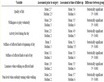
Table 1. qPCR primer information
表1. qPCR引物信息
2.8. Western Blot
用含有蛋白酶抑制剂和磷酸酶抑制剂的放射免疫共沉淀测定(RIPA)裂解缓冲液在冰上裂解6孔板中的细胞10分钟。收集上清液,用BCA试剂盒测定蛋白质样品的浓度。各蛋白样品经SDS-PAGE分离,电印迹到PVDF膜上。然后用一抗封闭,4℃孵育过夜。洗膜后加入二抗室温孵育1 h。采用ECL显影液于显影仪显影。用Image J软件进行密度分析。
2.9. 统计学
本实验结果数据利用Graphpad Prism 10.0软件进行统计分析和作图。正态分布的数据利用两独立样本T检验或单因素方差分析,非正态分布的数据利用Wilcoxon秩和检验及Kruskal-Wallis秩和检验。P < 0.05代表差异有统计学意义。
3. 结果
3.1. PBC细胞模型建立
PBC的病理特征是HiBECs凋亡性损伤,HiBECs凋亡后,PDC-E2暴露诱导胆管上皮产生自身免疫损伤的靶抗原。因此,需要检测HiBECs凋亡率和PDC-E2的表达水平,来验证PBC细胞模型是否构建成功。采用1 nM的GCDC作用于HiBECs 12 h后,采用qPCR法检测PDC-E2 mRNA相对表达量,结果发现PDC-E2 mRNA水平相较于NC组表达升高约2.3倍(P < 0.05) (见图1(a)),Western Blot结果显示PDC-E2蛋白表达水平升高约2.1倍(P < 0.01) (见图1(b)、图1(c))。同时采用流式细胞术检测细胞凋亡百分情况,结果显示正常的早期凋亡细胞在12.01%,而应用了GCDC诱导后的细胞凋亡率明显增加,达到31.78% (见图1(d))。以上证明了PBC细胞模型成功建立,可以用于后续研究。
 (a) 正常HiBECs组和GCDC组PDC-E2 mRNA相对表达水平;(b) 正常HiBECs组和GCDC组PDC-E2蛋白表达水平;(c) PDC-E2蛋白的定量分析;(d) 流式细胞术检测HiBECs凋亡率。
Figure 1. Establishment of the PBC cell model
图1. PBC细胞模型的建立
3.2. 1,25(OH)2D3对正常HiBEC细胞增殖活性的影响
为筛选出对HiBECs合适浓度,采用CCK-8法检测不同浓度1,25(OH)2D3对正常HiBECs增殖水平的影响。不同浓度(1 nM、10 nM、100 nM、300 nM) 1,25(OH)2D3处理正常HiBECs 12 h,实验结果表明,当1,25(OH)2D3浓度达到300 nM时,HiBECs增殖活性受到明显抑制(P < 0.05) (见图2(a)),而采用1 nM、10 nM、100 nM 1,25(OH)2D3处理对HiBECs增殖活性无明显抑制作用(P > 0.05) (见图3(a))。因此,本研究后续研究中,分别以1 nM、10 nM、100 nM作为维生素D的低、中、高浓度(GCDC + VD-L、GCDC + VD-M、GCDC + VD-H)进行实验。
3.3. 1,25(OH)2D3对PBC细胞分泌细胞因子的影响
我们通过ELISA检测细胞培养液中的IL-6、IL-8促炎细胞因子,结果发现,与NC组相比较,应用GCDC诱导的PBC细胞的培养液中促炎细胞因子IL-6 (P < 0.01) (见图3(a))、IL-8 (P < 0.05) (见图3(b))浓度均显著升高。应用1,25(OH)2D3处理后,GCDC + VD-L组、GCDC + VD-M和GCDC + VD-H组IL-6浓度均呈浓度梯度性下降(见图3(a))。与GCDC组相比,GCDC + VD-H组的IL-8浓度出现下降(P < 0.05) (见图3(b))。
 (a) 1,25(OH)2D3对正常HiBECs增殖活性的影响;(b) 1,25(OH)2D3对PBC细胞增殖活性的影响。#P < 0.05, ##P < 0.01 vs. NC组. *P < 0.05, **P < 0.01, ***P < 0.001 vs. GCDC组。ΔP < 0.05 vs. GCDC + VD-L组。GCDC + VD-L组:GCDC + 1 nM 1,25(OH)2D3;GCDC + VD-M组:GCDC + 10 nM 1,25(OH)2D3;GCDC + VD-H组:GCDC + 100 nM 1,25(OH)2D3。
Figure 2. The effect of 1,25(OH)2D3 on the proliferation of HiBECs
图2. 1,25(OH)2D3对HiBECs增殖的影响
 (a) 各组培养基中IL-6水平;(b) 各组培养液中IL-8水平。#P < 0.05, ##P < 0.01 vs. NC组。*P < 0.05, **P < 0.01, ***P < 0.001 vs. GCDC组。ΔP < 0.05 vs. GCDC + VD-L组。GCDC + VD-L组:GCDC + 1 nM 1,25(OH)2D3;GCDC + VD-M组:GCDC + 10 nM 1,25(OH)2D3;GCDC + VD-H组:GCDC + 100 nM 1,25(OH)2D3。
Figure 3. The effect of 1,25(OH)2D3 on IL-6 and IL-8 in cell culture fluid
图3. 1,25(OH)2D3对细胞培养液中IL-6、IL-8的影响
3.4. 1,25(OH)2D3对PBC细胞凋亡率的影响
早期凋亡细胞为膜联蛋白V-FITC阳性和PI阴性。如图5(a)所示,与对照组相比,GCDC组早期细胞凋亡增加,而维生素处理部分逆转了GCDC诱导的HIBEC凋亡,差异具有统计学意义(P < 0.05) (见图4(a))。检测凋亡相关标志物的表达。结果发现,与对照组相比,PDC-E2 mRNA及蛋白表达上升,而Bcl-2蛋白表达下调。与GCDC组相比,应用1,25(OH)2D3后的PDC-E2 mRNA及蛋白表达水平下降,而Bcl-2蛋白表达上调(见图4(b)、图4(c))。
3.5. 1,25(OH)2D3对细胞mtDNA的影响
COX1用于测定mtDNA拷贝数,PGC-1α、NRF1、NRF2和TFAM作为参与线粒体生物合成的转录

(a) 维生素D对HiBECs凋亡率的影响;(b) 维生素D对PBC细胞增殖活性的影响;(c) 各组培养基中IL-6水平;(d) PDC-E2蛋白的定量分析;(e) Bcl-2蛋白的定量分析。#P < 0.05, ##P < 0.01 vs. NC组。*P < 0.05, **P < 0.01, ***P < 0.001 vs. GCDC组。ΔP < 0.05 vs. GCDC + VD-L组。GCDC + VD-L组:GCDC + 1 nM 1,25(OH)2D3;GCDC + VD-M组:GCDC + 10 nM 1,25(OH)2D3;GCDC + VD-H组:GCDC + 100 nM 1,25(OH)2D3。
图4. 1,25(OH)2D3对HiBECs细胞凋亡率的影响
因子,是mtDNA增殖标志物。通过qPCR检测,结果发现与NC组相比,GCDC的处理显著降低了PGC-1α (P < 0.01,见图5(a))、Nrf-1 (P < 0.01) (见图5(b)),Nrf-2 (P < 0.01) (见图5(c))和TFAM (P < 0.05) (见图5(d))、COX1 (P < 0.01) (见图5(e))的mRNA水平。而1,25(OH)2D3的干预逆转了GCDC的负面影响,使Nrf-1 (P < 0.01) (图5(b)),Nrf-2 (P < 0.01) (图5(b))和TFAM (P < 0.05) (图5(d))、COX1 (P < 0.05) (图5(e)) mRNA表达明显升高。

(a) qPCR检测各组COX1 mRNA表达水平。(b) qPCR检测各组PGC-1α mRNA表达水平。(c) qPCR检测各组Nrf-1 mRNA表达水平。(d) qPCR检测各组Nrf-2 mRNA表达水平。(e) qPCR检测各组TFAM mRNA水平。#P < 0.05,##P < 0.01 vs. NC组。*P < 0.05,**P < 0.01,***P < 0.001 vs. GCDC组。ΔP < 0.05 vs. GCDC + VD-L组。GCDC + VD-L组:GCDC + 1 nM 1,25(OH)2D3;GCDC + VD-M组:GCDC + 10 nM 1,25(OH)2D3;GCDC + VD-H组:GCDC + 100 nM 1,25(OH)2D3。
图5. 1,25(OH)2D3对GCDC诱导的HiBECs mtDNA的影响
4. 讨论
在本研究中,我们建立了GCDC诱导的PBC细胞模型,以检测1,25(OH)2D3在PBC中的功能。近年来的研究表明维生素D对PBC有保护作用 [25] [26] [27] 。然而,大多数研究主要集中在血清中维生素D水平与PBC疾病进展的关系上,少数研究探讨了1,25(OH)2D3在PBC细胞模型中的影响及其机制。本研究首次发现1,25(OH)2D3可通过稳定mtDNA,调节线粒体代谢抑制GCDC诱导的HiBECs凋亡,可能是治疗PBC的潜在靶点。
首先本研究选择了人胆汁内主要毒性成分GCDC诱导人肝内胆管上皮细胞凋亡,PBC特异性自身抗体PDC-E2表达升高,细胞凋亡率增加,证明我们成功构建了PBC细胞模型,并且通过CCK-8实验确定了维生素D的合适作用剂量范围。实验结果显示GCDC诱导的HiBEC细胞增殖活性显著降低,细胞培养液中炎症因子IL-6、IL-8水平升高,同时HiBEC凋亡水平也显著增加。而维生素D提高了GCDC诱导的HiBEC细胞增殖活性,降低了细胞培养液中IL-6和IL-8水平,并且可以通过稳定mtDNA影响线粒体功能,抑制GCDC诱导的HiBEC凋亡,说明了维生素D对GCDC诱导的HiBEC具有保护作用。。这些结果提示维生素D可能是治疗PBC的一种选择。
以往的一些研究发现,维生素D可抑制细胞凋亡,在多种疾病中发挥治疗和预防作用。维生素D能减少顺铂诱导的人肾小管上皮细胞凋亡 [28] ,保护胃黏膜上皮细胞免受幽门螺旋杆菌诱导的凋亡 [29] ,改善链脲佐菌素诱导的大鼠胰腺组织凋亡和氧化应激 [30] 。维生素D对慢性肝胆疾病 [31] [32] [33] ,尤其是自身免疫性肝病(AILD)有保护作用。自身免疫性肝病(AILD)患者血清维生素D水平普遍降低,补充血清维生素D可减轻肝损伤和肝纤维化程度 [27] [34] [35] 。与之前研究结果一致,本研究发现维生素D减少PBC的HiBEC凋亡,在PBC中发挥保护作用。
线粒体功能障碍与肝胆汁淤积症有关。线粒体功能障碍与肝胆汁淤积症有关 [36] 。有毒胆盐积累会导致慢性损伤,如线粒体损伤、ROS增加和细胞凋亡,从而导致肝功能异常。研究发现,PGC-1α在维持线粒体和能量代谢平衡方面具有重要功能 [36] 。PGC-1α主导线粒体DNA复制和细胞氧化代谢,其表达水平是线粒体基因表达的限制因素 [37] 。此外,PGC-1α与NrFs (如NrF-1和NrF-2)共同作用,提高TFAM的表达来控制线粒体的生物生成 [38] [39] 。迄今为止,很少有研究报道1,25(OH)2D3通过线粒体途径对细胞凋亡具有保护作用。而我们的研究也发现,GCDC处理HiBEC通过抑制线粒体DNA稳定性、降低PGC-1α表达诱导细胞凋亡。维生素D通过激活PGC-1α,增加NrF-1,NrF-2表达,稳定mtDNA,并减少促炎细胞因子,减少HiBEC的细胞凋亡。
综上所述,本研究表明GCDC诱导HiBECs凋亡,1,25(OH)2D3通过稳定mtDNA途径参与GCDC诱导的HiBECs的抗凋亡作用。我们的研究结果阐明了1,25(OH)2D3在PBC中发挥保护作用的新机制。然而,该研究仍有一些不可忽视的局限性。目前的研究仅限于体外细胞模型,需要进一步的动物模型和临床实验,以全面阐明维生素D对PBC的治疗作用和机制,为PBC的研究提供理论基础。
5. 结论
本研究结果表明,维生素D可通过减轻线粒体mtDNA损伤,来抑制GCDC诱导的HiBEC凋亡,最终对PBC具有保护作用。这些结果拓展了我们对维生素D在PBC中治疗的认识,并为治疗PBC提供了新的靶点思路。
作者贡献声明
李永新负责文章撰写、实验设计及数据整理;王战负责实验验证、数据分析整理;高炜泽参与数据整理和实验验证;鲁文龙参与数据收集;刘明军负责课题总体设计和文章修改。
利益冲突
本人与其他作者宣称没有任何利益冲突,未接受任何不当的职务或财务利益。
文章引用
李永新,王 战,高炜泽,鲁文龙,刘明军. 1,25(OH)2D3通过稳定线粒体DNA对GCDC诱导的HiBECs凋亡的作用研究
The Role of 1,25(OH)2D3 on GCDC-Induced Apoptosis in HiBECs by Stabilising Mitochondrial DNA[J]. 临床医学进展, 2024, 14(04): 2256-2266. https://doi.org/10.12677/acm.2024.1441290
参考文献
- 1. Carey, E.J., Ali, A.H. and Lindor, K.D. (2015) Primary Biliary Cirrhosis. The Lancet, 386, 1565-1575.https://doi.org/10.1016/S0140-6736(15)00154-3
- 2. Laschtowitz, A., De Veer, R.C., Van Der, Meer, A.J., et al. (2020) Diagnosis and Treatment of Primary Biliary Cholangitis. United European Gastroenterology Journal, 8, 667-674. https://doi.org/10.1177/2050640620919585
- 3. Li, H., Guan, Y., Han, C., Zhang, Y., Liu, Q., Wei, W. and Ma, Y. (2021) The Pathogenesis, Models and Therapeutic Advances of Primary Biliary Cholangitis. Biomedicine & Pharmacotherapy, 140, Article 111754. https://pubmed.ncbi.nlm.nih.gov/34044277/https://doi.org/10.1016/j.biopha.2021.111754
- 4. Spiechowicz, M., Zylicz, A., Bieganowski, P., et al. (2007) Hsp70 Is a New Target of Sgt1—An Interaction Modulated by S100A6. Biochemical and Biophysical Research Communications, 357, 1148-1153.https://doi.org/10.1016/j.bbrc.2007.04.073
- 5. Rust, C., Wild, N., Bernt, C., et al. (2009) Bile Acid-Induced Apoptosis in Hepatocytes Is Caspase-6-Dependent. The Journal of Biological Chemistry, 284, 2908-2916. https://doi.org/10.1074/jbc.M804585200
- 6. Arduini, A., Serviddio, G., Escobar, J., et al. (2011) Mitochondrial Biogenesis Fails in Secondary Biliary Cirrhosis in Rats Leading to Mitochondrial DNA Depletion and Deletions. American Journal of Physiology-Gastrointestinal and Liver Physiology, 301, G119-G127. https://doi.org/10.1152/ajpgi.00253.2010
- 7. Pejznochova, M., Tesarova, M., Hansikova, H., et al. (2010) Mitochondrial DNA Content and Expression of Genes Involved in mtDNA Transcription, Regulation and Maintenance during Human Fetal Development. Mitochondrion, 10, 321-329. https://doi.org/10.1016/j.mito.2010.01.006
- 8. Heidari, R. and Niknahad, H. (2019) The Role and Study of Mitochondrial Impairment and Oxidative Stress in Cholestasis. In: Vinken, M., Ed., Experimental Cholestasis Research. Methods in Molecular Biology, Vol. 1981, Humana, New York, 117-132. https://doi.org/10.1007/978-1-4939-9420-5_8
- 9. Picca, A., Calvani, R., Coelho-Junior, H.J., et al. (2021) Cell Death and Inflammation: The Role of Mitochondria in Health and Disease. Cells, 10, Article 537. https://doi.org/10.3390/cells10030537
- 10. Alexeyev, M., Shokolenko, I., Wilson, G. and LeDoux, S. (2013) The Maintenance of Mitochondrial DNA Integrity—Critical Analysis and Update. Cold Spring Harbor Perspectives in Biology, 5, Article a012641.https://pubmed.ncbi.nlm.nih.gov/23637283/https://doi.org/10.1101/cshperspect.a012641
- 11. Saki, M. and Prakash, A. (2017) DNA Damage Related Crosstalk between the Nucleus and Mitochondria. Free Radical Biology and Medicine, 107, 216-227. https://doi.org/10.1016/j.freeradbiomed.2016.11.050
- 12. Bouillon, R., Marcocci, C., Carmeliet, G., et al. (2019) Skeletal and Extraskeletal Actions of Vitamin D: Current Evidence and Outstanding Questions. Endocrine Reviews, 40, 1109-1151. https://doi.org/10.1210/er.2018-00126
- 13. Fleet, J.C. and Schoch, R.D. (2010) Molecular Mechanisms for Regulation of Intestinal Calcium Absorption by Vitamin D and Other Factors. Critical Reviews in Clinical Laboratory Sciences, 47, 181-195.https://doi.org/10.3109/10408363.2010.536429
- 14. Walker, V.P. and Modlin, R.L. (2009) The Vitamin D Connection to Pediatric Infections and Immune Function. Pediatric Research, 65, 106-113. https://doi.org/10.1203/PDR.0b013e31819dba91
- 15. Holick, M.F. (2007) Vitamin D Deficiency. The New England Journal of Medicine, 357, 266-281.https://doi.org/10.1056/NEJMra070553
- 16. Plum, L.A. and DeLuca, H.F. (2010) Vitamin D, Disease and Therapeutic Opportunities. Nature Reviews Drug Discovery, 9, 941-955. https://doi.org/10.1038/nrd3318
- 17. Kempinska-Podhorodecka, A., Milkiewicz, M., Wasik, U., et al. (2017) Decreased Expression of Vitamin D Receptor Affects an Immune Response in Primary Biliary Cholangitis via the VDR-miRNA155-SOCS1 Pathway. International Journal of Molecular Sciences, 18, Article 289. https://doi.org/10.3390/ijms18020289
- 18. Banerjee, A., Athalye, S., Khargekar, N., et al. (2023) Chronic Hepatitis B and Related Liver Diseases Are Associated with Reduced 25-Hydroxy-Vitamin D Levels: A Systematic Review and Meta-Analysis. Biomedicines, 11, Article 135.https://doi.org/10.3390/biomedicines11010135
- 19. Chan, H.L.-Y., Elkhashab, M., Trinh, H., et al. (2015) Association of Baseline Vitamin D Levels with Clinical Parameters and Treatment Outcomes in Chronic Hepatitis B. Journal of Hepatology, 63, 1086-1092.https://doi.org/10.1016/j.jhep.2015.06.025
- 20. García-Álvarez, M., Pineda-Tenor, D., Jiménez-Sousa, M.A., et al. (2014) Relationship of Vitamin D Status with Advanced Liver Fibrosis and Response to Hepatitis C Virus Therapy: A Meta-Analysis. Hepatology, 60, 1541-1550.https://doi.org/10.1002/hep.27281
- 21. Udomsinprasert, W., Jittikoon, J., Sukkho, S., et al. (2020) Decreased Circulating Vitamin D Reflects Adverse Outcomes of Hepatitis C Virus Infection: A Systematic Review and Meta-Analysis. The Journal of Infection, 81, 585-599.https://doi.org/10.1016/j.jinf.2020.06.025
- 22. Tao, S., Zhang, H., Zhao, Q., et al. (2020) Correlation of Vitamin D with Inflammatory Factors, Oxidative Stress and T Cell Subsets in Patients with Autoimmune Hepatitis. Experimental and Therapeutic Medicine, 19, 3419-3424.https://doi.org/10.3892/etm.2020.8601
- 23. Reda, D., Elshopakey, G.E., Albukhari, T.A., et al. (2023) Vitamin D3 Alleviates Nonalcoholic Fatty Liver Disease in Rats by Inhibiting Hepatic Oxidative Stress and Inflammation via the SREBP-1-C/ PPARα-NF-κB/IR-S2 Signaling Pathway. Frontiers in Pharmacology, 14, Article 1164512. https://doi.org/10.3389/fphar.2023.1164512
- 24. Stepan, M.D., Vintilescu Ș.B., Streață, I., et al. (2023) The Role of Vitamin D in Obese Children with Non-Alcoholic Fatty Liver Disease and Associated Metabolic Syndrome. Nutrients, 15, Article 2113.https://doi.org/10.3390/nu15092113
- 25. Xu, H., Wu, Z., Feng, F., et al. (2022) Low Vitamin D Concentrations and BMI Are Causal Factors for Primary Biliary Cholangitis: A Mendelian Randomization Study. Frontiers in Immunology, 13, Article 1055953.https://doi.org/10.3389/fimmu.2022.1055953
- 26. Wang, Z., Peng, C., Wang, P., et al. (2020) Serum Vitamin D Level Is Related to Disease Progression in Primary Biliary Cholangitis. Scandinavian Journal of Gastroenterology, 55, 1333-1340.https://doi.org/10.1080/00365521.2020.1829030
- 27. Ebadi, M., Ip, S., Lytvyak, E., et al. (2022) Vitamin D Is Associated with Clinical Outcomes in Patients with Primary Biliary Cholangitis. Nutrients, 14, Article 878. https://doi.org/10.3390/nu14040878
- 28. Jiang, S., Zhang, H., Li, X., et al. (2021) Vitamin D/VDR Attenuate Cisplatin-Induced AKI by Down-Regulating NLRP3/Caspase-1/GSDMD Pyroptosis Pathway. The Journal of Steroid Biochemistry and Molecular Biology, 206, Article 105789. https://doi.org/10.1016/j.jsbmb.2020.105789
- 29. Zhao, S., Zhong, Y., Xu, X. and Wan, D. (2022) 1α, 25-Dihydroxyvitamin D3 Protects Gastric Mucosa Epithelial Cells against Helicobacter pylori-Infected Apoptosis through a Vitamin D Receptor-Dependent c-Raf/MEK/ERK Pathway. Pharmaceutical Biology, 60, 801-809. https://doi.org/10.1080/13880209.2022.2058559
- 30. Fathi, F.E.Z.M., Sadek, K.M., Khafaga, A.F., et al. (2022) Correction to: Vitamin D Regulates Insulin and Ameliorates Apoptosis and Oxidative Stress in Pancreatic Tissues of Rats with Streptozotocin-Induced Diabetes. Environmental Science and Pollution Research International, 29, 90230. https://doi.org/10.1007/s11356-022-22427-9
- 31. Grünhage, F., Hochrath, K., Krawczyk, M., et al. (2012) Common Genetic Variation in Vitamin D Metabolism Is Associated with Liver Stiffness. Hepatology, 56, 1883-1891. https://doi.org/10.1002/hep.25830
- 32. Petta, S., Cammà, C., Scazzone, C., et al. (2010) Low Vitamin D Serum Level Is Related to Severe Fibrosis and Low Responsiveness to Interferon-Based Therapy in Genotype 1 Chronic Hepatitis C. Hepatology, 51, 1158-1167.https://doi.org/10.1002/hep.23489
- 33. Tourkochristou, E., Tsounis, E.P., Tzoupis, H., et al. (2023) The Influence of Single Nucleotide Polymorphisms on Vitamin D Receptor Protein Levels and Function in Chronic Liver Disease. International Journal of Molecular Sciences, 24, Article 11404. https://doi.org/10.3390/ijms241411404
- 34. Ebadi, M., Rider, E., Tsai, C., et al. (2023) Prognostic Significance of Severe Vitamin D Deficiency in Patients with Primary Sclerosing Cholangitis. Nutrients, 15, Article 576. https://doi.org/10.3390/nu15030576
- 35. Hochrath, K., Stokes, C.S., Geisel, J., et al. (2014) Vitamin D Modulates Biliary Fibrosis in ABCB4-Deficient Mice. Hepatology International, 8, 443-452. https://doi.org/10.1007/s12072-014-9548-2
- 36. Mihaylov, S.R., Castelli, L.M., Lin, Y.-H., et al. (2023) The Master Energy Homeostasis Regulator PGC-1α Exhibits an MRNA Nuclear Export Function. Nature Communications, 14, Article No. 5496.https://doi.org/10.1038/s41467-023-41304-8
- 37. Rubalcava-Gracia, D., García-Villegas, R. and Larsson, N.-G. (2023) No Role for Nuclear Transcription Regulators in Mammalian Mitochondria? Molecular Cell, 83, 832-842. https://doi.org/10.1016/j.molcel.2022.09.010
- 38. Ongwijitwat, S., Liang, H.L., Graboyes, E.M., et al. (2006) Nuclear Respiratory Factor 2 Senses Changing Cellular Energy Demands and Its Silencing Down-Regulates Cytochrome Oxidase and Other Target Gene mRNAs. Gene, 374, 39-49. https://doi.org/10.1016/j.gene.2006.01.009
- 39. Scarpulla, R.C. (2008) Transcriptional Paradigms in Mammalian Mitochondrial Biogenesis and Function. Physiological Reviews, 88, 611-638. https://doi.org/10.1152/physrev.00025.2007
NOTES
*通讯作者。